A monovalent cation acts as structural and catalytic cofactor in translational GTPases.
Kuhle, B., Ficner, R.(2014) EMBO J 33: 2547-2563
- PubMed: 25225612 Search on PubMedSearch on PubMed Central
- DOI: https://doi.org/10.15252/embj.201488517
- Primary Citation Related Structures:
4TMT, 4TMV, 4TMW, 4TMX, 4TMZ, 4TN1 - PubMed Abstract:
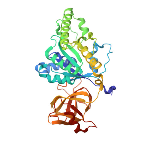
Translational GTPases are universally conserved GTP hydrolyzing enzymes, critical for fidelity and speed of ribosomal protein biosynthesis. Despite their central roles, the mechanisms of GTP-dependent conformational switching and GTP hydrolysis that govern the function of trGTPases remain poorly understood. Here, we provide biochemical and high-resolution structural evidence that eIF5B and aEF1A/EF-Tu bound to GTP or GTPγS coordinate a monovalent cation (M(+)) in their active site. Our data reveal that M(+) ions form constitutive components of the catalytic machinery in trGTPases acting as structural cofactor to stabilize the GTP-bound "on" state. Additionally, the M(+) ion provides a positive charge into the active site analogous to the arginine-finger in the Ras-RasGAP system indicating a similar role as catalytic element that stabilizes the transition state of the hydrolysis reaction. In sequence and structure, the coordination shell for the M(+) ion is, with exception of eIF2γ, highly conserved among trGTPases from bacteria to human. We therefore propose a universal mechanism of M(+)-dependent conformational switching and GTP hydrolysis among trGTPases with important consequences for the interpretation of available biochemical and structural data.
- Abteilung für Molekulare Strukturbiologie, Institut für Mikrobiologie und Genetik Göttinger Zentrum für Molekulare Biowissenschaften Georg-August-Universität Göttingen, Göttingen, Germany bkuhle@gwdg.de.
Organizational Affiliation: