Structural basis for efficient phosphorylation of 3'-azidothymidine monophosphate by Escherichia coli thymidylate kinase.
Lavie, A., Ostermann, N., Brundiers, R., Goody, R.S., Reinstein, J., Konrad, M., Schlichting, I.(1998) Proc Natl Acad Sci U S A 95: 14045-14050
- PubMed: 9826650 Search on PubMedSearch on PubMed Central
- DOI: https://doi.org/10.1073/pnas.95.24.14045
- Primary Citation Related Structures:
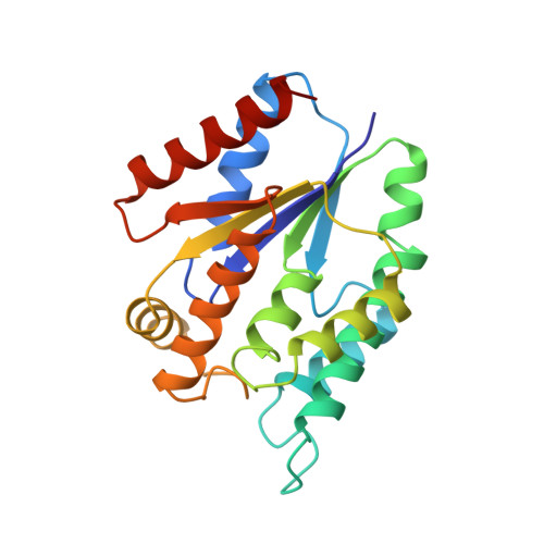
4TMK, 5TMP - PubMed Abstract:
The crystal structures of Escherichia coli thymidylate kinase (TmpK) in complex with P1-(5'-adenosyl)-P5-(5'-thymidyl)pentaphosphate and P1-(5'-adenosyl)P5-[5'-(3'-azido-3'-deoxythymidine)] pentaphosphate have been solved to 2.0-A and 2.2-A resolution, respectively. The overall structure of the bacterial TmpK is very similar to that of yeast TmpK. In contrast to the human and yeast TmpKs, which phosphorylate 3'-azido-3'-deoxythymidine 5'-monophosphate (AZT-MP) at a 200-fold reduced turnover number (kcat) in comparison to the physiological substrate dTMP, reduction of kcat is only 2-fold for the bacterial enzyme. The different kinetic properties toward AZT-MP between the eukaryotic TmpKs and E. coli TmpK can be rationalized by the different ways in which these enzymes stabilize the presumed transition state and the different manner in which a carboxylic acid side chain in the P loop interacts with the deoxyribose of the monophosphate. Yeast TmpK interacts with the 3'-hydroxyl of dTMP through Asp-14 of the P loop in a bidentate manner: binding of AZT-MP results in a shift of the P loop to accommodate the larger substituent. In E. coli TmpK, the corresponding residue is Glu-12, and it interacts in a side-on fashion with the 3'-hydroxyl of dTMP. This different mode of interaction between the P loop carboxylic acid with the 3' substituent of the monophosphate deoxyribose allows the accommodation of an azido group in the case of the E. coli enzyme without significant P loop movement. In addition, although the yeast enzyme uses Arg-15 (a glycine in E. coli) to stabilize the transition state, E. coli seems to use Arg-153 from a region termed Lid instead. Thus, the binding of AZT-MP to the yeast TmpK results in the shift of a catalytic residue, which is not the case for the bacterial kinase.
- Department of Physical Biochemistry, Max Planck Institute for Molecular Physiology, Rheinlanddamm 201, 44139 Dortmund, Germany.
Organizational Affiliation: