Structure-based Conversion of the Coenzyme Requirement of a Short-chain Dehydrogenase/Reductase Involved in Bacterial Alginate Metabolism.
Takase, R., Mikami, B., Kawai, S., Murata, K., Hashimoto, W.(2014) J Biol Chem 289: 33198-33214
- PubMed: 25288804 Search on PubMedSearch on PubMed Central
- DOI: https://doi.org/10.1074/jbc.M114.585661
- Primary Citation Related Structures:
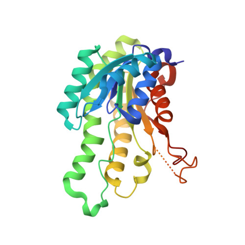
4TKL, 4TKM, 4W7H, 4W7I - PubMed Abstract:
The alginate-assimilating bacterium, Sphingomonas sp. strain A1, degrades the polysaccharides to monosaccharides through four alginate lyase reactions. The resultant monosaccharide, which is nonenzymatically converted to 4-deoxy-L-erythro-5-hexoseulose uronate (DEH), is further metabolized to 2-keto-3-deoxy-D-gluconate by NADPH-dependent reductase A1-R in the short-chain dehydrogenase/reductase (SDR) family. A1-R-deficient cells produced another DEH reductase, designated A1-R', with a preference for NADH. Here, we show the identification of a novel NADH-dependent DEH reductase A1-R' in strain A1, structural determination of A1-R' by x-ray crystallography, and structure-based conversion of a coenzyme requirement in SDR enzymes, A1-R and A1-R'. A1-R' was purified from strain A1 cells and enzymatically characterized. Except for the coenzyme requirement, there was no significant difference in enzyme characteristics between A1-R and A1-R'. Crystal structures of A1-R' and A1-R'·NAD(+) complex were determined at 1.8 and 2.7 Å resolutions, respectively. Because of a 64% sequence identity, overall structures of A1-R' and A1-R were similar, although a difference in the coenzyme-binding site (particularly the nucleoside ribose 2' region) was observed. Distinct from A1-R, A1-R' included a negatively charged, shallower binding site. These differences were caused by amino acid residues on the two loops around the site. The A1-R' mutant with the two A1-R-typed loops maintained potent enzyme activity with specificity for NADPH rather than NADH, demonstrating that the two loops determine the coenzyme requirement, and loop exchange is a promising method for conversion of coenzyme requirement in the SDR family.
- From the Laboratory of Basic and Applied Molecular Biotechnology, Division of Food Science and Biotechnology, and.
Organizational Affiliation: