


Crystal Structure of nucleoside diphosphate kinase at 1.92 A resolution from Acinetobacter baumannii
Sikarwar, J., Shukla, P.K., Kaur, P., Sharma, S., Singh, T.P.To be published.
Experimental Data Snapshot
Starting Model: experimental
View more details
wwPDB Validation 3D Report Full Report

Entity ID: 1 | |||||
|---|---|---|---|---|---|
| Molecule | Chains | Sequence Length | Organism | Details | Image |
| Nucleoside diphosphate kinase | 143 | Acinetobacter baumannii | Mutation(s): 0 Gene Names: ABBL099_02780, BL01_17035, FL75_03050, FL80_07750, GQ86_04945, IOMTU433_3298, IX87_17090, LX00_02580, ndk, P795_14800 EC: 2.7.4.6 | 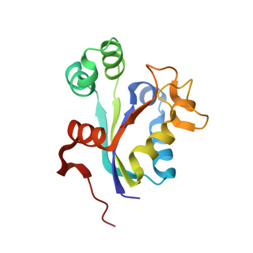 | |
UniProt | |||||
Entity Groups | |||||
| Sequence Clusters | 30% Identity50% Identity70% Identity90% Identity95% Identity100% Identity | ||||
| UniProt Group | V5VIC4 | ||||
Sequence AnnotationsExpand | |||||
Reference Sequence | |||||
| Ligands 1 Unique | |||||
|---|---|---|---|---|---|
| ID | Chains | Name / Formula / InChI Key | 2D Diagram | 3D Interactions | |
| MG Download:Ideal Coordinates CCD File | I [auth D], J [auth G] | MAGNESIUM ION Mg JLVVSXFLKOJNIY-UHFFFAOYSA-N |  | ||
| Length ( Å ) | Angle ( ˚ ) |
|---|---|
| a = 68.924 | α = 90 |
| b = 71.381 | β = 93.71 |
| c = 129.427 | γ = 90 |
| Software Name | Purpose |
|---|---|
| HKL-2000 | data collection |
| AMoRE | phasing |
| PHENIX | refinement |
| HKL-2000 | data reduction |
| SCALEPACK | data scaling |


