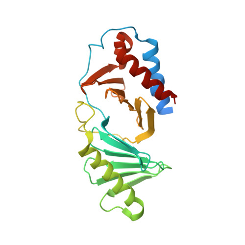
4RS6

Crystal structure of the C domain of Polo like Kinase II in Homo Sapiens


Experimental Data Snapshot

  • Method: X-RAY DIFFRACTION
  • Resolution: 2.60 Å
  • R-Value Free: 0.247 
  • R-Value Work: 0.186 
  • R-Value Observed: 0.189 

Starting Model: experimental
View more details

wwPDB Validation   3D Report Full Report


This is version 1.2 of the entry. See complete history


Literature

Crystal structure of the polo-box domain of polo-like kinase 2

Shan, H.M.Wang, T.Quan, J.M.

(2015) Biochem Biophys Res Commun 456: 780-784

  • DOI: https://doi.org/10.1016/j.bbrc.2014.11.125
  • Primary Citation of Related Structures:  
    4RS6

  • PubMed Abstract: 

    Polo-like kinase 2 (PLK2) is a crucial regulator in cell cycle progression, DNA damage response, and neuronal activity. PLK2 is characterized by the conserved N-terminal kinase domain and the unique C-terminal polo-box domain (PBD). The PBD mediates diverse functions of PLK2 by binding phosphorylated Ser-pSer/pThr motifs of its substrates. Here, we report the first crystal structure of the PBD of PLK2. The overall structure of the PLK2 PBD is similar to that of the PLK1 PBD, which is composed by two polo boxes each contain β6α structures that form a 12-stranded β sandwich domain. The edge of the interface between the two polo boxes forms the phosphorylated Ser-pSer/pThr motifs binding cleft. On the hand, the peripheral regions around the core binding cleft of the PLK2 PBD is distinct from that of the PLK1 PBD, which might confer the substrate specificity of the PBDs of the polo-like kinase family.


  • Organizational Affiliation

    Key Laboratory of Structural Biology, School of Chemical Biology & Biotechnology, Peking University Shenzhen Graduate School, Shenzhen 518055, China.


Macromolecules
Find similar proteins by:  (by identity cutoff)  |  3D Structure
Entity ID: 1
MoleculeChains Sequence LengthOrganismDetailsImage
Serine/threonine-protein kinase PLK2
A, B
242Homo sapiensMutation(s): 0 
Gene Names: PLK2SNK
EC: 2.7.11.21
UniProt & NIH Common Fund Data Resources
Find proteins for Q9NYY3 (Homo sapiens)
Explore Q9NYY3 
Go to UniProtKB:  Q9NYY3
PHAROS:  Q9NYY3
GTEx:  ENSG00000145632 
Entity Groups  
Sequence Clusters30% Identity50% Identity70% Identity90% Identity95% Identity100% Identity
UniProt GroupQ9NYY3
Sequence Annotations
Expand
  • Reference Sequence
Experimental Data & Validation

Experimental Data

  • Method: X-RAY DIFFRACTION
  • Resolution: 2.60 Å
  • R-Value Free: 0.247 
  • R-Value Work: 0.186 
  • R-Value Observed: 0.189 
  • Space Group: I 2 3
Unit Cell:
Length ( Å )Angle ( ˚ )
a = 153.075α = 90
b = 153.075β = 90
c = 153.075γ = 90
Software Package:
Software NamePurpose
Blu-Icedata collection
PHENIXmodel building
REFMACrefinement
HKL-2000data reduction
HKL-2000data scaling
PHENIXphasing

Structure Validation

View Full Validation Report


Entry History 

Deposition Data

Revision History  (Full details and data files)

  • Version 1.0: 2014-12-03
    Type: Initial release
  • Version 1.1: 2016-06-08
    Changes: Database references
  • Version 1.2: 2023-11-08
    Changes: Data collection, Database references, Refinement description