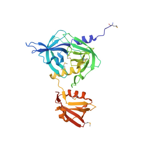
4RR1

re-refinement of entry 1sot, Crystal Structure of the DegS stress sensor


Experimental Data Snapshot

  • Method: X-RAY DIFFRACTION
  • Resolution: 2.30 Å
  • R-Value Free: 0.217 
  • R-Value Work: 0.184 
  • R-Value Observed: 0.186 

wwPDB Validation   3D Report Full Report


This is version 1.2 of the entry. See complete history

Re-refinement Note

This entry reflects an alternative modeling of the original data in: 1SOT


Literature

A Conserved Activation Cluster Is Required for Allosteric Communication in HtrA-Family Proteases.

de Regt, A.K.Kim, S.Sohn, J.Grant, R.A.Baker, T.A.Sauer, R.T.

(2015) Structure 23: 517-526

  • DOI: https://doi.org/10.1016/j.str.2015.01.012
  • Primary Citation of Related Structures:  
    4RQY, 4RQZ, 4RR0, 4RR1

  • PubMed Abstract: 

    In E. coli, outer-membrane stress causes a transcriptional response through a signaling cascade initiated by DegS cleavage of a transmembrane antisigma factor. Each subunit of DegS, an HtrA-family protease, contains a protease domain and a PDZ domain. The trimeric protease domain is autoinhibited by the unliganded PDZ domains. Allosteric activation requires binding of unassembled outer-membrane proteins (OMPs) to the PDZ domains and protein substrate binding. Here, we identify a set of DegS residues that cluster together at subunit-subunit interfaces in the trimer, link the active sites and substrate binding sites, and are crucial for stabilizing the active enzyme conformation in response to OMP signaling. These residues are conserved across the HtrA-protease family, including orthologs linked to human disease, supporting a common mechanism of allosteric activation. Indeed, mutation of residues at homologous positions in the DegP quality-control protease also eliminates allosteric activation.


  • Organizational Affiliation

    Department of Biology, Massachusetts Institute of Technology, 31 Ames Street, Cambridge, MA 02139, USA.


Macromolecules
Find similar proteins by:  (by identity cutoff)  |  3D Structure
Entity ID: 1
MoleculeChains Sequence LengthOrganismDetailsImage
Protease degS
A, B, C
320Escherichia coliMutation(s): 0 
Gene Names: degSP12B_c3347
EC: 3.4.21.107
UniProt
Find proteins for P0AEE3 (Escherichia coli (strain K12))
Explore P0AEE3 
Go to UniProtKB:  P0AEE3
Entity Groups  
Sequence Clusters30% Identity50% Identity70% Identity90% Identity95% Identity100% Identity
UniProt GroupP0AEE3
Sequence Annotations
Expand
  • Reference Sequence
Small Molecules
Modified Residues  1 Unique
IDChains TypeFormula2D DiagramParent
MSE
Query on MSE
A, B, C
L-PEPTIDE LINKINGC5 H11 N O2 SeMET
Experimental Data & Validation

Experimental Data

  • Method: X-RAY DIFFRACTION
  • Resolution: 2.30 Å
  • R-Value Free: 0.217 
  • R-Value Work: 0.184 
  • R-Value Observed: 0.186 
  • Space Group: C 1 2 1
Unit Cell:
Length ( Å )Angle ( ˚ )
a = 207.68α = 90
b = 143.06β = 90.07
c = 41.53γ = 90
Software Package:
Software NamePurpose
PHENIXrefinement
PDB_EXTRACTdata extraction

Structure Validation

View Full Validation Report


Entry History 

Deposition Data

Revision History  (Full details and data files)

  • Version 1.0: 2015-03-11
    Type: Initial release
  • Version 1.1: 2015-03-18
    Changes: Database references
  • Version 1.2: 2024-11-27
    Changes: Data collection, Database references, Derived calculations, Structure summary