4RQS | pdb_00004rqs

Crystal structure of fully glycosylated HIV-1 gp120 core bound to CD4 and 17b Fab


Experimental Data Snapshot

  • Method: X-RAY DIFFRACTION
  • Resolution: 4.49 Å
  • R-Value Free: 
    0.322 (Depositor), 0.339 (DCC) 
  • R-Value Work: 
    0.276 (Depositor), 0.283 (DCC) 
  • R-Value Observed: 
    0.279 (Depositor) 

wwPDB Validation 3D Report Full Report

Validation slider image for 4RQS

Ligand Structure Quality Assessment 


This is version 2.1 of the entry. See complete history

Literature

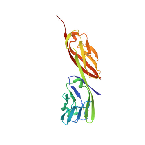
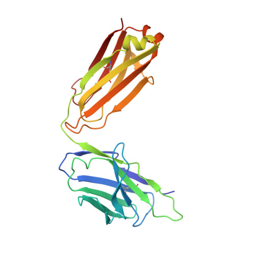
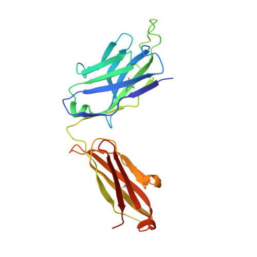
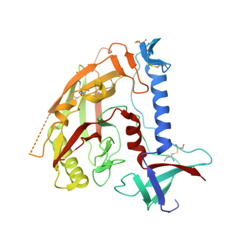
Crystal structure of a fully glycosylated HIV-1 gp120 core reveals a stabilizing role for the glycan at Asn262.

Kong, L.Wilson, I.A.Kwong, P.D.

(2015) Proteins 83: 590-596

  • DOI: https://doi.org/10.1002/prot.24747
  • Primary Citation Related Structures: 
    4RQS

  • PubMed Abstract: 

    The crystal structure of a fully glycosylated HIV-1 gp120 core in complex with CD4 receptor and Fab 17b at 4.5-Å resolution reveals 9 of the 15 N-linked glycans of core gp120 to be partially ordered. The glycan at position Asn262 had the most extensive and well-ordered electron density, and a GlcNAc(2)Man(7) was modeled. The GlcNAc stem of this glycan is largely buried in a cleft in gp120, suggesting a role in gp120 folding and stability. Its arms interact with the stems of neighboring glycans from the oligomannose patch, which is a major target for broadly neutralizing antibodies.


  • Organizational Affiliation
    • Vaccine Research Center, National Institutes of Health, Bethesda, Maryland, 20892; Department of Integrative Structural and Computational Biology, the Scripps Research Institute, La Jolla, California; International AIDS Vaccine Initiative Neutralizing Antibody Center and the Collaboration for AIDS Vaccine Discovery, the Scripps Research Institute, La Jolla, California; Scripps Center for HIV/AIDS Vaccine Immunology and Immunogen Discovery, the Scripps Research Institute, La Jolla, California.

Macromolecule Content 

  • Total Structure Weight: 108.44 kDa 
  • Atom Count: 7,379 
  • Modeled Residue Count: 913 
  • Deposited Residue Count: 941 
  • Unique protein chains: 4

Macromolecules

Find similar proteins by:|  3D Structure
Entity ID: 1
MoleculeChains  Sequence LengthOrganismDetailsImage
2-domain CD4A [auth B]185Homo sapiensMutation(s): 0 
UniProt & NIH Common Fund Data Resources
Find proteins for P01730 (Homo sapiens)
Explore P01730 
Go to UniProtKB:  P01730
PHAROS:  P01730
GTEx:  ENSG00000010610 
Entity Groups
Sequence Clusters30% Identity50% Identity70% Identity90% Identity95% Identity100% Identity
UniProt GroupP01730
Sequence Annotations
Expand
Reference Sequence
Find similar proteins by:|  3D Structure
Entity ID: 2
MoleculeChains  Sequence LengthOrganismDetailsImage
17b Fab Light ChainB [auth C]214Homo sapiensMutation(s): 0 
Find similar proteins by:|  3D Structure
Entity ID: 3
MoleculeChains  Sequence LengthOrganismDetailsImage
17b Fab Heavy ChainC [auth D]229Homo sapiensMutation(s): 0 
Find similar proteins by:|  3D Structure
Entity ID: 4
MoleculeChains  Sequence LengthOrganismDetailsImage
HIV-1 YU2 gp120 core chimeric proteinD [auth G]313Human immunodeficiency virus type 1 (isolate YU2)Mutation(s): 0 
UniProt
Find proteins for P35961 (Human immunodeficiency virus type 1 group M subtype B (isolate YU-2))
Explore P35961 
Go to UniProtKB:  P35961
Entity Groups
Sequence Clusters30% Identity50% Identity70% Identity90% Identity95% Identity100% Identity
UniProt GroupP35961
Glycosylation
Glycosylation Sites: 9
Sequence Annotations
Expand
Reference Sequence

Oligosaccharides

Help  
Entity ID: 5
MoleculeChains Length2D Diagram GlycosylationD Interactions
alpha-D-mannopyranose-(1-2)-alpha-D-mannopyranose-(1-2)-alpha-D-mannopyranose-(1-3)-[alpha-D-mannopyranose-(1-3)-[alpha-D-mannopyranose-(1-6)]alpha-D-mannopyranose-(1-6)]beta-D-mannopyranose-(1-4)-2-acetamido-2-deoxy-beta-D-glucopyranose-(1-4)-2-acetamido-2-deoxy-beta-D-glucopyranoseE [auth A]9N-Glycosylation
Glycosylation Resources
GlyTouCan: G68668TB
GlyCosmos: G68668TB
GlyGen: G68668TB
Entity ID: 6
MoleculeChains Length2D Diagram GlycosylationD Interactions
2-acetamido-2-deoxy-beta-D-glucopyranose-(1-4)-2-acetamido-2-deoxy-beta-D-glucopyranoseF [auth E],
H,
I,
J
2N-Glycosylation
Glycosylation Resources
GlyTouCan: G42666HT
GlyCosmos: G42666HT
GlyGen: G42666HT
Entity ID: 7
MoleculeChains Length2D Diagram GlycosylationD Interactions
beta-D-mannopyranose-(1-4)-2-acetamido-2-deoxy-beta-D-glucopyranose-(1-4)-2-acetamido-2-deoxy-beta-D-glucopyranoseG [auth F],
K,
L
3N-Glycosylation
Glycosylation Resources
GlyTouCan: G15407YE
GlyCosmos: G15407YE
GlyGen: G15407YE

Small Molecules

Ligands 1 Unique
IDChains Name / Formula / InChI Key2D Diagram3D Interactions
NAG

Query on NAG


Download:Ideal Coordinates CCD File
M [auth G]2-acetamido-2-deoxy-beta-D-glucopyranose
C8 H15 N O6
OVRNDRQMDRJTHS-FMDGEEDCSA-N

Experimental Data & Validation

Experimental Data

  • Method: X-RAY DIFFRACTION
  • Resolution: 4.49 Å
  • R-Value Free:  0.322 (Depositor), 0.339 (DCC) 
  • R-Value Work:  0.276 (Depositor), 0.283 (DCC) 
  • R-Value Observed: 0.279 (Depositor) 
Space Group: P 43 21 2
Unit Cell:
Length ( Å )Angle ( ˚ )
a = 171.318α = 90
b = 171.318β = 90
c = 151.857γ = 90
Software Package:
Software NamePurpose
Blu-Icedata collection
PHASERphasing
PHENIXrefinement
HKL-2000data reduction
SCALEPACKdata scaling

Structure Validation

View Full Validation Report


Ligand Structure Quality Assessment 


Entry History 

Deposition Data

Revision History  (Full details and data files)

  • Version 1.0: 2014-12-31
    Type: Initial release
  • Version 1.1: 2015-01-14
    Changes: Database references
  • Version 1.2: 2015-02-25
    Changes: Database references
  • Version 1.3: 2017-06-28
    Changes: Source and taxonomy
  • Version 2.0: 2020-07-29
    Type: Remediation
    Reason: Carbohydrate remediation
    Changes: Advisory, Atomic model, Data collection, Database references, Derived calculations, Structure summary
  • Version 2.1: 2024-10-09
    Changes: Data collection, Database references, Structure summary