4RPB | pdb_00004rpb

Crystal Structure of P Domain of Snow Mountain Norovirus


Experimental Data Snapshot

  • Method: X-RAY DIFFRACTION
  • Resolution: 1.61 Å
  • R-Value Free: 
    0.182 (Depositor), 0.175 (DCC) 
  • R-Value Work: 
    0.165 (Depositor) 
  • R-Value Observed: 
    0.166 (Depositor) 

wwPDB Validation 3D Report Full Report

Validation slider image for 4RPB

This is version 2.1 of the entry. See complete history

Literature

Structural Constraints on Human Norovirus Binding to Histo-Blood Group Antigens.

Singh, B.K.Leuthold, M.M.Hansman, G.S.

(2016) mSphere 1

  • DOI: https://doi.org/10.1128/mSphere.00049-16
  • Primary Citation Related Structures: 
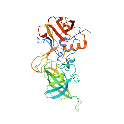
    4ROX, 4RPB, 4RPD

  • PubMed Abstract: 

    Human norovirus interacts with the polymorphic human histo-blood group antigens (HBGAs), and this interaction is thought to be important for infection. The genogroup II genotype 4 (GII.4) noroviruses are the dominant cluster, evolve every other year, and are thought to modify their binding interactions with different HBGA types. Most human noroviruses bind HBGAs, while some strains were found to have minimal or no HBGA interactions. Here, we explain some possible structural constraints for several noroviruses that were found to bind poorly to HBGAs by using X-ray crystallography. We showed that one aspartic acid was flexible or positioned away from the fucose moiety of the HBGAs and this likely hindered binding, although other fucose-interacting residues were perfectly oriented. Interestingly, a neighboring loop also appeared to influence the loop hosting the aspartic acid. These new findings might explain why some human noroviruses bound HBGAs poorly, although further studies are required.


  • Organizational Affiliation
    • Schaller Research Group at the University of Heidelberg and the DKFZ, Heidelberg, Germany; Department of Infectious Diseases, Virology, University of Heidelberg, Heidelberg, Germany.

Macromolecule Content 

  • Total Structure Weight: 102.47 kDa 
  • Atom Count: 7,641 
  • Modeled Residue Count: 900 
  • Deposited Residue Count: 930 
  • Unique protein chains: 1

Macromolecules

Find similar proteins by:|  3D Structure
Entity ID: 1
MoleculeChains  Sequence LengthOrganismDetailsImage
Capsid protein VP1
A, B, C
310Norovirus Hu/GII.2/KL109/1978/MYSMutation(s): 0 
Gene Names: VP1
UniProt
Find proteins for K7X601 (Norovirus Hu/GII.2/KL109/1978/MYS)
Explore K7X601 
Go to UniProtKB:  K7X601
Entity Groups
Sequence Clusters30% Identity50% Identity70% Identity90% Identity95% Identity100% Identity
UniProt GroupK7X601
Sequence Annotations
Expand
Reference Sequence

Experimental Data & Validation

Experimental Data

  • Method: X-RAY DIFFRACTION
  • Resolution: 1.61 Å
  • R-Value Free:  0.182 (Depositor), 0.175 (DCC) 
  • R-Value Work:  0.165 (Depositor) 
  • R-Value Observed: 0.166 (Depositor) 
Space Group: C 1 2 1
Unit Cell:
Length ( Å )Angle ( ˚ )
a = 166.64α = 90
b = 96.17β = 90
c = 64.12γ = 90
Software Package:
Software NamePurpose
ADSCdata collection
PHASERphasing
PHENIXrefinement
XDSdata reduction
XDSdata scaling

Structure Validation

View Full Validation Report


Entry History 

Deposition Data

Revision History  (Full details and data files)

  • Version 1.0: 2016-06-22
    Type: Initial release
  • Version 1.1: 2016-06-29
    Changes: Database references
  • Version 2.0: 2022-08-24
    Changes: Atomic model, Database references, Derived calculations
  • Version 2.1: 2024-02-28
    Changes: Data collection