


Crystal structure of esterase A from Streptococcus pyogenes
Bennett, M.D., Holland, R., Coulibaly, F., Loo, T.S., Norris, G.E., Anderson, B.F.To be published.
Experimental Data Snapshot
wwPDB Validation 3D Report Full Report

Entity ID: 1 | |||||
|---|---|---|---|---|---|
| Molecule | Chains | Sequence Length | Organism | Details | Image |
| Esterase A | 268 | Streptococcus pyogenes | Mutation(s): 1 Gene Names: Est A, HMPREF1245_1265 EC: 3.1.1.1 (PDB Primary Data), 3.1.1.2 (PDB Primary Data) | 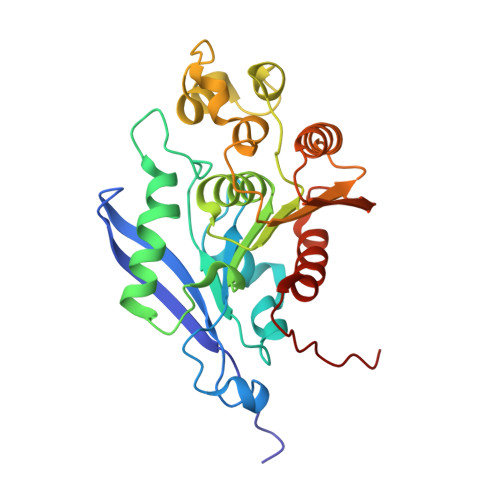 | |
| Ligands 2 Unique | |||||
|---|---|---|---|---|---|
| ID | Chains | Name / Formula / InChI Key | 2D Diagram | 3D Interactions | |
| OXL Download:Ideal Coordinates CCD File | C [auth A] | OXALATE ION C2 O4 MUBZPKHOEPUJKR-UHFFFAOYSA-L |  | ||
| ZN Download:Ideal Coordinates CCD File | B [auth A] | ZINC ION Zn PTFCDOFLOPIGGS-UHFFFAOYSA-N |  | ||
| Length ( Å ) | Angle ( ˚ ) |
|---|---|
| a = 103.98 | α = 90 |
| b = 103.98 | β = 90 |
| c = 133.631 | γ = 90 |
| Software Name | Purpose |
|---|---|
| ADSC | data collection |
| SOLVE | phasing |
| REFMAC | refinement |
| DENZO | data reduction |
| SCALEPACK | data scaling |


