4RL3 | pdb_00004rl3

Crystal Structure of the Catalytic Domain of a family GH18 Chitinase from fern, Peteris ryukyuensis


Experimental Data Snapshot

  • Method: X-RAY DIFFRACTION
  • Resolution: 1.57 Å
  • R-Value Free: 
    0.165 (Depositor), 0.178 (DCC) 
  • R-Value Work: 
    0.150 (Depositor), 0.165 (DCC) 
  • R-Value Observed: 
    0.151 (Depositor) 

Starting Model: experimental
View more details

wwPDB Validation 3D Report Full Report

Validation slider image for 4RL3

This is version 1.1 of the entry. See complete history

Literature

A class III chitinase without disulfide bonds from the fern, Pteris ryukyuensis: crystal structure and ligand-binding studies.

Kitaoku, Y.Umemoto, N.Ohnuma, T.Numata, T.Taira, T.Sakuda, S.Fukamizo, T.

(2015) Planta 242: 895-907

  • DOI: https://doi.org/10.1007/s00425-015-2330-4
  • Primary Citation Related Structures: 
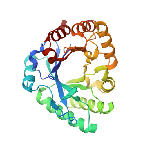
    4RL3

  • PubMed Abstract: 

    We first solved the crystal structure of class III catalytic domain of a chitinase from fern (PrChiA-cat), and found a structural difference between PrChiA-cat and hevamine. PrChiA-cat was found to have reduced affinities to chitin oligosaccharides and allosamidin. Plant class III chitinases are subdivided into enzymes with three disulfide bonds and those without disulfide bonds. We here referred to the former enzymes as class IIIa chitinases and the latter as class IIIb chitinases. In this study, we solved the crystal structure of the class IIIb catalytic domain of a chitinase from the fern Pteris ryukyuensis (PrChiA-cat), and compared it with that of hevamine, a class IIIa chitinase from Hevea brasiliensis. PrChiA-cat was found to adopt an (α/β)8 fold typical of GH18 chitinases in a similar manner to that of hevamine. However, PrChiA-cat also had two large loops that extruded from the catalytic site, and the corresponding loops in hevamine were markedly smaller than those of PrChiA-cat. An HPLC analysis of the enzymatic products revealed that the mode of action of PrChiA-cat toward chitin oligosaccharides, (GlcNAc) n (n = 4-6), differed from those of hevamine and the other class IIIa chitinases. The binding affinities of (GlcNAc)3 and (GlcNAc)4 toward the inactive mutant of PrChiA-cat were determined by isothermal titration calorimetry, and were markedly lower than those toward other members of the GH18 family. The affinity and the inhibitory activity of allosamidin toward PrChiA-cat were also lower than those toward the GH18 chitinases investigated to date. Several hydrogen bonds found in the crystal structure of hevamine-allosamidin complex were missing in the modeled structure of PrChiA-cat-allosamidin complex. The structural findings for PrChiA-cat successfully interpreted the functional data presented.


  • Organizational Affiliation
    • Department of Advanced Bioscience, Kinki University, 3327-204 Nakamachi, Nara, 631-8505, Japan.

Macromolecule Content 

  • Total Structure Weight: 57.96 kDa 
  • Atom Count: 4,637 
  • Modeled Residue Count: 542 
  • Deposited Residue Count: 544 
  • Unique protein chains: 1

Macromolecules

Find similar proteins by:|  3D Structure
Entity ID: 1
MoleculeChains  Sequence LengthOrganismDetailsImage
Chitinase A
A, B
272Pteris ryukyuensisMutation(s): 0 
Gene Names: prchiA
EC: 3.2.1.14
UniProt
Find proteins for Q0WYK2 (Pteris ryukyuensis)
Explore Q0WYK2 
Go to UniProtKB:  Q0WYK2
Entity Groups
Sequence Clusters30% Identity50% Identity70% Identity90% Identity95% Identity100% Identity
UniProt GroupQ0WYK2
Sequence Annotations
Expand
Reference Sequence

Small Molecules

Ligands 3 Unique
IDChains Name / Formula / InChI Key2D Diagram3D Interactions
EDO

Query on EDO


Download:Ideal Coordinates CCD File
C [auth A],
D [auth A],
G [auth B],
H [auth B]
1,2-ETHANEDIOL
C2 H6 O2
LYCAIKOWRPUZTN-UHFFFAOYSA-N
ACT

Query on ACT


Download:Ideal Coordinates CCD File
E [auth A],
I [auth B]
ACETATE ION
C2 H3 O2
QTBSBXVTEAMEQO-UHFFFAOYSA-M
MG

Query on MG


Download:Ideal Coordinates CCD File
F [auth A],
J [auth B]
MAGNESIUM ION
Mg
JLVVSXFLKOJNIY-UHFFFAOYSA-N

Experimental Data & Validation

Experimental Data

  • Method: X-RAY DIFFRACTION
  • Resolution: 1.57 Å
  • R-Value Free:  0.165 (Depositor), 0.178 (DCC) 
  • R-Value Work:  0.150 (Depositor), 0.165 (DCC) 
  • R-Value Observed: 0.151 (Depositor) 
Space Group: I 41
Unit Cell:
Length ( Å )Angle ( ˚ )
a = 78.626α = 90
b = 78.626β = 90
c = 167.4γ = 90
Software Package:
Software NamePurpose
HKL-2000data collection
MOLREPphasing
REFMACrefinement
HKL-2000data reduction
HKL-2000data scaling

Structure Validation

View Full Validation Report


Entry History 

Deposition Data

Revision History  (Full details and data files)

  • Version 1.0: 2015-10-14
    Type: Initial release
  • Version 1.1: 2023-11-08
    Changes: Data collection, Database references, Derived calculations, Refinement description