Opposing orientations of the anti-psychotic drug trifluoperazine selected by alternate conformations of M144 in calmodulin.
Feldkamp, M.D., Gakhar, L., Pandey, N., Shea, M.A.(2015) Proteins 83: 989-996
- PubMed: 25694384 Search on PubMedSearch on PubMed Central
- DOI: https://doi.org/10.1002/prot.24781
- Primary Citation Related Structures:
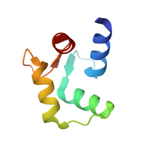
4RJD - PubMed Abstract:
The anti-psychotic drug trifluoperazine (TFP) is an antagonist observed to bind to calcium-saturated calmodulin ((Ca(2+) )4 -CaM) at ratios of 1:1 (1CTR), 2:1 (1A29), and 4:1 (1LIN). Each structure contains one TFP bound in the hydrophobic cleft of the C-domain of CaM. However, the orientation of the trifluoromethyl (CF3 ) moiety differs among them: it is buried in the C-domain cleft of 1A29 and 1LIN, but protrudes from 1CTR. We report a 2.0 Å resolution crystallographic structure (4RJD) of TFP bound to the (Ca(2+) )-saturated C-domain of CaM (CaMC ). The asymmetric unit contains two molecules of (Ca(2+) )2 -CaMC . Chain backbones were nearly identical, but the orientation of TFP in the cleft of Chain A matched 1A29/1LIN, while TFP bound to Chain B matched 1CTR. This was accommodated by a flip of the M144 sidechain and small changes in sidechains of M109 and M145. Docking simulations suggested that the rotamer conformation of M144 determined the orientation of TFP within the cleft of (Ca(2+) )2 -CaMC . Chains A and B show that the open cleft of (Ca(2+) )2 -CaMC is promiscuous in accepting TFP in reversed directions under the same crystallization conditions. Observing multiple orientations of an antagonist bound to a single protein highlights the challenge of designing highly specific pharmaceuticals, and may have importance for QSAR of other CF3 -containing drugs such as fluoxetine (anti-depressant) or efavirenz (reverse transcriptase inhibitor). This study emphasizes that a single structure of a complex represents an energetically accessible state, but does not necessarily show the full range of energetically equivalent states.
- Department of Biochemistry, Roy J. and Lucille A. Carver College of Medicine, University of Iowa, Iowa City, Iowa, 52242-1109.
Organizational Affiliation: