4RG2

Tudor Domain of Tumor suppressor p53BP1 with small molecule ligand


Experimental Data Snapshot

  • Method: X-RAY DIFFRACTION
  • Resolution: 1.50 Å
  • R-Value Free: 0.220 
  • R-Value Work: 0.197 
  • R-Value Observed: 0.198 

wwPDB Validation   3D Report Full Report


Ligand Structure Quality Assessment 


This is version 1.3 of the entry. See complete history


Literature

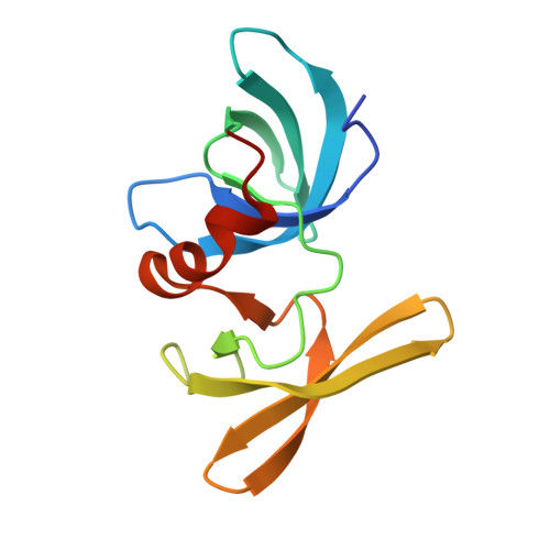
Identification of a fragment-like small molecule ligand for the methyl-lysine binding protein, 53BP1.

Perfetti, M.T.Baughman, B.M.Dickson, B.M.Mu, Y.Cui, G.Mader, P.Dong, A.Norris, J.L.Rothbart, S.B.Strahl, B.D.Brown, P.J.Janzen, W.P.Arrowsmith, C.H.Mer, G.McBride, K.M.James, L.I.Frye, S.V.

(2015) ACS Chem Biol 10: 1072-1081

  • DOI: https://doi.org/10.1021/cb500956g
  • Primary Citation of Related Structures:  
    4RG2

  • PubMed Abstract: 

    Improving our understanding of the role of chromatin regulators in the initiation, development, and suppression of cancer and other devastating diseases is critical, as they are integral players in regulating DNA integrity and gene expression. Developing small molecule inhibitors for this target class with cellular activity is a crucial step toward elucidating their specific functions. We specifically targeted the DNA damage response protein, 53BP1, which uses its tandem tudor domain to recognize histone H4 dimethylated on lysine 20 (H4K20me2), a modification related to double-strand DNA breaks. Through a cross-screening approach, we identified UNC2170 (1) as a micromolar ligand of 53BP1, which demonstrates at least 17-fold selectivity for 53BP1 as compared to other methyl-lysine (Kme) binding proteins tested. Structural studies revealed that the tert-butyl amine of UNC2170 anchors the compound in the methyl-lysine (Kme) binding pocket of 53BP1, making it competitive with endogenous Kme substrates. X-ray crystallography also demonstrated that UNC2170 binds at the interface of two tudor domains of a 53BP1 dimer. Importantly, this compound functions as a 53BP1 antagonist in cellular lysates and shows cellular activity by suppressing class switch recombination, a process which requires a functional 53BP1 tudor domain. These results demonstrate that UNC2170 is a functionally active, fragment-like ligand for 53BP1.


  • Organizational Affiliation

    †Center for Integrative Chemical Biology and Drug Discovery, Division of Chemical Biology and Medicinal Chemistry, UNC Eshelman School of Pharmacy, University of North Carolina at Chapel Hill, Chapel Hill, North Carolina 27599, United States.


Macromolecules
Find similar proteins by:  (by identity cutoff)  |  3D Structure
Entity ID: 1
MoleculeChains Sequence LengthOrganismDetailsImage
Tumor suppressor p53-binding protein 1
A, B
125Homo sapiensMutation(s): 0 
Gene Names: TP53BP1
UniProt & NIH Common Fund Data Resources
Find proteins for Q12888 (Homo sapiens)
Explore Q12888 
Go to UniProtKB:  Q12888
PHAROS:  Q12888
GTEx:  ENSG00000067369 
Entity Groups  
Sequence Clusters30% Identity50% Identity70% Identity90% Identity95% Identity100% Identity
UniProt GroupQ12888
Sequence Annotations
Expand
  • Reference Sequence
Small Molecules
Ligands 3 Unique
IDChains Name / Formula / InChI Key2D Diagram3D Interactions
3OO
Query on 3OO

Download Ideal Coordinates CCD File 
C [auth A]3-bromo-N-[3-(tert-butylamino)propyl]benzamide
C14 H21 Br N2 O
XUFUVCIZFNREBI-UHFFFAOYSA-N
EDO
Query on EDO

Download Ideal Coordinates CCD File 
D [auth A],
E [auth A],
M [auth B]
1,2-ETHANEDIOL
C2 H6 O2
LYCAIKOWRPUZTN-UHFFFAOYSA-N
UNX
Query on UNX

Download Ideal Coordinates CCD File 
F [auth A]
G [auth A]
H [auth A]
I [auth A]
J [auth A]
F [auth A],
G [auth A],
H [auth A],
I [auth A],
J [auth A],
K [auth A],
L [auth A],
N [auth B],
O [auth B],
P [auth B],
Q [auth B],
R [auth B]
UNKNOWN ATOM OR ION
X
Binding Affinity Annotations 
IDSourceBinding Affinity
3OO BindingDB:  4RG2 Kd: 2.20e+4 (nM) from 1 assay(s)
IC50: min: 2.90e+4, max: 3.00e+4 (nM) from 2 assay(s)
Experimental Data & Validation

Experimental Data

  • Method: X-RAY DIFFRACTION
  • Resolution: 1.50 Å
  • R-Value Free: 0.220 
  • R-Value Work: 0.197 
  • R-Value Observed: 0.198 
  • Space Group: C 2 2 21
Unit Cell:
Length ( Å )Angle ( ˚ )
a = 73.548α = 90
b = 100.458β = 90
c = 83.348γ = 90
Software Package:
Software NamePurpose
DENZOdata reduction
SCALEPACKdata scaling
PHASERphasing
REFMACrefinement
PDB_EXTRACTdata extraction
SBC-Collectdata collection
HKL-3000data reduction
HKL-3000data scaling
Cootmodel building
GRADErefinement

Structure Validation

View Full Validation Report


Ligand Structure Quality Assessment 


Entry History 

Revision History  (Full details and data files)

  • Version 1.0: 2014-10-15
    Type: Initial release
  • Version 1.1: 2017-11-22
    Changes: Refinement description
  • Version 1.2: 2018-05-16
    Changes: Data collection, Database references
  • Version 1.3: 2023-09-20
    Changes: Data collection, Database references, Derived calculations, Refinement description