4REA

A Nuclease DNA complex


Experimental Data Snapshot

  • Method: X-RAY DIFFRACTION
  • Resolution: 3.81 Å
  • R-Value Free: 0.277 
  • R-Value Work: 0.243 
  • R-Value Observed: 0.244 

wwPDB Validation   3D Report Full Report


This is version 1.2 of the entry. See complete history


Literature

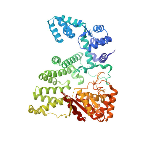
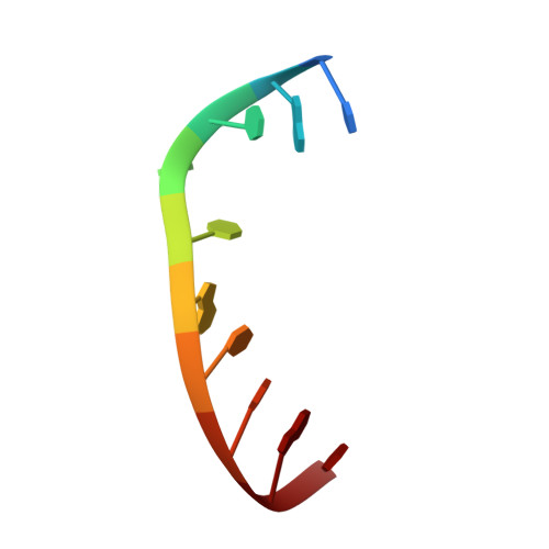
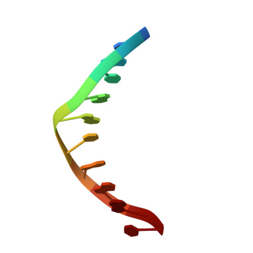
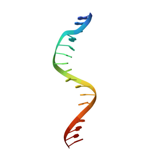
Structural insights into 5' flap DNA unwinding and incision by the human FAN1 dimer.

Zhao, Q.Xue, X.Longerich, S.Sung, P.Xiong, Y.

(2014) Nat Commun 5: 5726-5726

  • DOI: https://doi.org/10.1038/ncomms6726
  • Primary Citation of Related Structures:  
    4REA, 4REB, 4REC

  • PubMed Abstract: 

    Human FANCD2-associated nuclease 1 (FAN1) is a DNA structure-specific nuclease involved in the processing of DNA interstrand crosslinks (ICLs). FAN1 maintains genomic stability and prevents tissue decline in multiple organs, yet it confers ICL-induced anti-cancer drug resistance in several cancer subtypes. Here we report three crystal structures of human FAN1 in complex with a 5' flap DNA substrate, showing that two FAN1 molecules form a head-to-tail dimer to locate the lesion, orient the DNA and unwind a 5' flap for subsequent incision. Biochemical experiments further validate our model for FAN1 action, as structure-informed mutations that disrupt protein dimerization, substrate orientation or flap unwinding impair the structure-specific nuclease activity. Our work elucidates essential aspects of FAN1-DNA lesion recognition and a unique mechanism of incision. These structural insights shed light on the cellular mechanisms underlying organ degeneration protection and cancer drug resistance mediated by FAN1.


  • Organizational Affiliation

    Department of Molecular Biophysics and Biochemistry, Yale University School of Medicine, New Haven, Connecticut 06520, USA.


Macromolecules

Find similar proteins by:  (by identity cutoff)  |  3D Structure
Entity ID: 1
MoleculeChains Sequence LengthOrganismDetailsImage
Fanconi-associated nuclease 1A [auth B],
B [auth A]
647Homo sapiensMutation(s): 1 
Gene Names: FAN1KIAA1018MTMR15
EC: 3.1.21 (PDB Primary Data), 3.1.4.1 (PDB Primary Data)
UniProt & NIH Common Fund Data Resources
Find proteins for Q9Y2M0 (Homo sapiens)
Explore Q9Y2M0 
Go to UniProtKB:  Q9Y2M0
PHAROS:  Q9Y2M0
GTEx:  ENSG00000198690 
Entity Groups  
Sequence Clusters30% Identity50% Identity70% Identity90% Identity95% Identity100% Identity
UniProt GroupQ9Y2M0
Sequence Annotations
Expand
  • Reference Sequence

Find similar nucleic acids by:  Sequence   |   3D Structure  

Entity ID: 2
MoleculeChains LengthOrganismImage
DNA (5'-D(*TP*GP*CP*TP*CP*GP*CP*CP*AP*C)-3')10synthetic construct
Sequence Annotations
Expand
  • Reference Sequence

Find similar nucleic acids by:  Sequence   |   3D Structure  

Entity ID: 3
MoleculeChains LengthOrganismImage
DNA (5'-D(P*CP*GP*TP*GP*GP*CP*GP*AP*GP*C)-3')10synthetic construct
Sequence Annotations
Expand
  • Reference Sequence

Find similar nucleic acids by:  Sequence   |   3D Structure  

Entity ID: 4
MoleculeChains LengthOrganismImage
DNA (5'-D(P*GP*GP*CP*GP*AP*GP*CP*GP*CP*TP*CP*GP*CP*CP*AP*CP*G)-3')17synthetic construct
Sequence Annotations
Expand
  • Reference Sequence
Experimental Data & Validation

Experimental Data

  • Method: X-RAY DIFFRACTION
  • Resolution: 3.81 Å
  • R-Value Free: 0.277 
  • R-Value Work: 0.243 
  • R-Value Observed: 0.244 
  • Space Group: P 31
Unit Cell:
Length ( Å )Angle ( ˚ )
a = 100.96α = 90
b = 100.96β = 90
c = 115.71γ = 120
Software Package:
Software NamePurpose
REFMACrefinement
PDB_EXTRACTdata extraction
CBASSdata collection

Structure Validation

View Full Validation Report


Entry History 

Deposition Data

Revision History  (Full details and data files)

  • Version 1.0: 2014-12-24
    Type: Initial release
  • Version 1.1: 2015-01-14
    Changes: Database references
  • Version 1.2: 2024-02-28
    Changes: Data collection, Database references, Refinement description