Lead Optimization and Modulation of hERG Activity in a Series of Aminooxazoline Xanthene beta-Site Amyloid Precursor Protein Cleaving Enzyme (BACE1) Inhibitors.
Epstein, O., Bryan, M.C., Cheng, A.C., Derakhchan, K., Dineen, T.A., Hickman, D., Hua, Z., Human, J.B., Kreiman, C., Marx, I.E., Weiss, M.M., Wahl, R.C., Wen, P.H., Whittington, D.A., Wood, S., Zheng, X.M., Fremeau, R.T., White, R.D., Patel, V.F.(2014) J Med Chem 57: 9796-9810
- PubMed: 25389560 Search on PubMed
- DOI: https://doi.org/10.1021/jm501266w
- Primary Citation Related Structures:
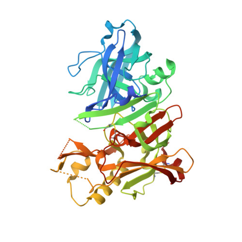
4RCE, 4RCF - PubMed Abstract:
The optimization of a series of aminooxazoline xanthene inhibitors of β-site amyloid precursor protein cleaving enzyme 1 (BACE1) is described. An early lead compound showed robust Aβ lowering activity in a rat pharmacodynamic model, but advancement was precluded by a low therapeutic window to QTc prolongation in cardiovascular models consistent with in vitro activity on the hERG ion channel. While the introduction of polar groups was effective in reducing hERG binding affinity, this came at the expense of higher than desired Pgp-mediated efflux. A balance of low Pgp efflux and hERG activity was achieved by lowering the polar surface area of the P3 substituent while retaining polarity in the P2' side chain. The introduction of a fluorine in position 4 of the xanthene ring improved BACE1 potency (5-10-fold). The combination of these optimized fragments resulted in identification of compound 40, which showed robust Aβ reduction in a rat pharmacodynamic model (78% Aβ reduction in CSF at 10 mg/kg po) and also showed acceptable cardiovascular safety in vivo.
- Departments of Therapeutic Discovery, ‡Neuroscience, §Molecular Structure and Characterization, ∥Pharmacokinetics and Drug Metabolism, and ⊥Comparative Biology and Safety Sciences, Amgen Inc. , 360 Binney Street, Cambridge, Massachusetts 02142, One Amgen Center Drive, Thousand Oaks, California 91320, and 1120 Veterans Boulevard, South San Francisco, California 94080, United States.
Organizational Affiliation: