4R97 | pdb_00004r97

Crystal structure of the Fab fragment of KKO


Experimental Data Snapshot

  • Method: X-RAY DIFFRACTION
  • Resolution: 2.20 Å
  • R-Value Free: 
    0.262 (Depositor), 0.266 (DCC) 
  • R-Value Work: 
    0.200 (Depositor), 0.203 (DCC) 
  • R-Value Observed: 
    0.203 (Depositor) 

wwPDB Validation 3D Report Full Report

Validation slider image for 4R97

This is version 1.3 of the entry. See complete history

Literature

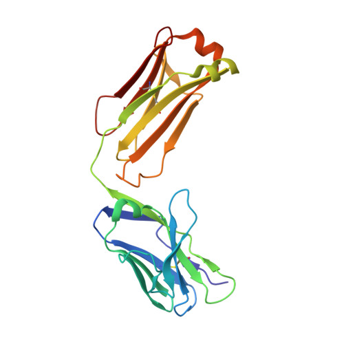
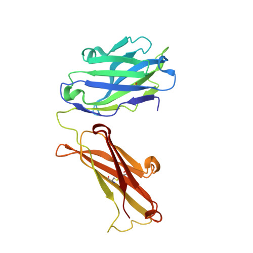
Atomic description of the immune complex involved in heparin-induced thrombocytopenia.

Cai, Z.Yarovoi, S.V.Zhu, Z.Rauova, L.Hayes, V.Lebedeva, T.Liu, Q.Poncz, M.Arepally, G.Cines, D.B.Greene, M.I.

(2015) Nat Commun 6: 8277-8277

  • DOI: https://doi.org/10.1038/ncomms9277
  • Primary Citation Related Structures: 
    4R97, 4R9W, 4R9Y

  • PubMed Abstract: 

    Heparin-induced thrombocytopenia (HIT) is an autoimmune thrombotic disorder caused by immune complexes containing platelet factor 4 (PF4), antibodies to PF4 and heparin or cellular glycosaminoglycans (GAGs). Here we solve the crystal structures of the: (1) PF4 tetramer/fondaparinux complex, (2) PF4 tetramer/KKO-Fab complex (a murine monoclonal HIT-like antibody) and (3) PF4 monomer/RTO-Fab complex (a non-HIT anti-PF4 monoclonal antibody). Fondaparinux binds to the 'closed' end of the PF4 tetramer and stabilizes its conformation. This interaction in turn stabilizes the epitope for KKO on the 'open' end of the tetramer. Fondaparinux and KKO thereby collaborate to 'stabilize' the ternary pathogenic immune complex. Binding of RTO to PF4 monomers prevents PF4 tetramerization and inhibits KKO and human HIT IgG-induced platelet activation and platelet aggregation in vitro, and thrombus progression in vivo. The atomic structures provide a basis to develop new diagnostics and non-anticoagulant therapeutics for HIT.


  • Organizational Affiliation
    • Department of Pathology and Laboratory Medicine, University of Pennsylvania, Philadelphia, Pennsylvania 19104, USA.

Macromolecule Content 

  • Total Structure Weight: 47.63 kDa 
  • Atom Count: 3,421 
  • Modeled Residue Count: 432 
  • Deposited Residue Count: 432 
  • Unique protein chains: 2

Macromolecules

Find similar proteins by:|  3D Structure
Entity ID: 1
MoleculeChains  Sequence LengthOrganismDetailsImage
platelet factor 4 antibody KKO light chainA [auth B]214Mus musculusMutation(s): 0 
Find similar proteins by:|  3D Structure
Entity ID: 2
MoleculeChains  Sequence LengthOrganismDetailsImage
platelet factor 4 antibody KKO heavy chainB [auth C]218Mus musculusMutation(s): 0 

Experimental Data & Validation

Experimental Data

  • Method: X-RAY DIFFRACTION
  • Resolution: 2.20 Å
  • R-Value Free:  0.262 (Depositor), 0.266 (DCC) 
  • R-Value Work:  0.200 (Depositor), 0.203 (DCC) 
  • R-Value Observed: 0.203 (Depositor) 
Space Group: P 21 21 21
Unit Cell:
Length ( Å )Angle ( ˚ )
a = 41.482α = 90
b = 92.124β = 90
c = 122.144γ = 90
Software Package:
Software NamePurpose
Blu-Icedata collection
MOLREPphasing
PHENIXrefinement
HKL-2000data reduction
HKL-2000data scaling

Structure Validation

View Full Validation Report


Entry History 

Deposition Data

Revision History  (Full details and data files)

  • Version 1.0: 2015-12-16
    Type: Initial release
  • Version 1.1: 2016-01-27
    Changes: Database references
  • Version 1.2: 2017-11-22
    Changes: Refinement description
  • Version 1.3: 2024-11-06
    Changes: Data collection, Database references, Derived calculations, Structure summary