


Crystal structure of 5-methylcytosine deaminase from Klebsiella pneumoniae liganded with 5-methylcytosine
Fedorov, A.A., Fedorov, E.V., Hitchcock, D.S., Raushel, F.M., Almo, S.C.To be published.
Experimental Data Snapshot
Starting Model: experimental
View more details
wwPDB Validation 3D Report Full Report

Entity ID: 1 | |||||
|---|---|---|---|---|---|
| Molecule | Chains | Sequence Length | Organism | Details | Image |
| Cytosine deaminase | 431 | Klebsiella pneumoniae 30660/NJST258_1 | Mutation(s): 0 Gene Names: KPNJ1_03949 EC: 3.5.4.1 | 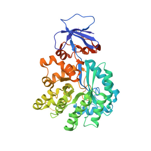 | |
Entity Groups | |||||
| Sequence Clusters | 30% Identity50% Identity70% Identity90% Identity95% Identity100% Identity | ||||
Sequence AnnotationsExpand | |||||
Reference Sequence | |||||
| Ligands 3 Unique | |||||
|---|---|---|---|---|---|
| ID | Chains | Name / Formula / InChI Key | 2D Diagram | 3D Interactions | |
| 17E Download:Ideal Coordinates CCD File | AA [auth E] G [auth A] GA [auth F] L [auth B] R [auth C] | 5-methylcytosine C5 H7 N3 O LRSASMSXMSNRBT-UHFFFAOYSA-N |  | ||
| GOL Download:Ideal Coordinates CCD File | CA [auth E] DA [auth E] EA [auth E] FA [auth F] I [auth A] | GLYCEROL C3 H8 O3 PEDCQBHIVMGVHV-UHFFFAOYSA-N |  | ||
| FE2 Download:Ideal Coordinates CCD File | BA [auth E] H [auth A] HA [auth F] M [auth B] S [auth C] | FE (II) ION Fe CWYNVVGOOAEACU-UHFFFAOYSA-N |  | ||
| Length ( Å ) | Angle ( ˚ ) |
|---|---|
| a = 111.481 | α = 90 |
| b = 137.669 | β = 118.03 |
| c = 112.093 | γ = 90 |
| Software Name | Purpose |
|---|---|
| CBASS | data collection |
| BALBES | phasing |
| PHENIX | refinement |
| HKL-2000 | data reduction |
| HKL-2000 | data scaling |


