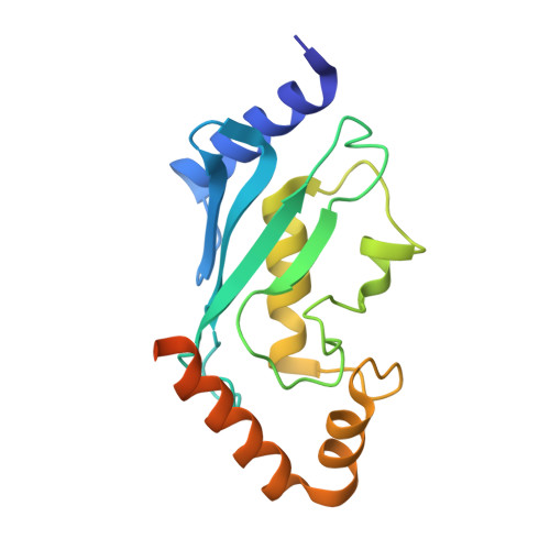
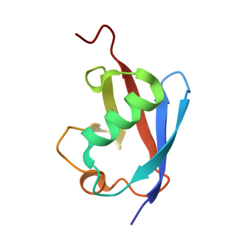
4R62

Structure of Rad6~Ub


Experimental Data Snapshot

  • Method: X-RAY DIFFRACTION
  • Resolution: 2.28 Å
  • R-Value Free: 0.265 
  • R-Value Work: 0.210 
  • R-Value Observed: 0.213 

Starting Models: experimental
View more details

wwPDB Validation   3D Report Full Report


This is version 1.2 of the entry. See complete history


Literature

Role of a non-canonical surface of Rad6 in ubiquitin conjugating activity.

Kumar, P.Magala, P.Geiger-Schuller, K.R.Majumdar, A.Tolman, J.R.Wolberger, C.

(2015) Nucleic Acids Res 43: 9039-9050

  • DOI: https://doi.org/10.1093/nar/gkv845
  • Primary Citation of Related Structures:  
    4R62

  • PubMed Abstract: 

    Rad6 is a yeast E2 ubiquitin conjugating enzyme that monoubiquitinates histone H2B in conjunction with the E3, Bre1, but can non-specifically modify histones on its own. We determined the crystal structure of a Rad6∼Ub thioester mimic, which revealed a network of interactions in the crystal in which the ubiquitin in one conjugate contacts Rad6 in another. The region of Rad6 contacted is located on the distal face of Rad6 opposite the active site, but differs from the canonical E2 backside that mediates free ubiquitin binding and polyubiquitination activity in other E2 enzymes. We find that free ubiquitin interacts weakly with both non-canonical and canonical backside residues of Rad6 and that mutations of non-canonical residues have deleterious effects on Rad6 activity comparable to those observed to mutations in the canonical E2 backside. The effect of non-canonical backside mutations is similar in the presence and absence of Bre1, indicating that contacts with non-canonical backside residues govern the intrinsic activity of Rad6. Our findings shed light on the determinants of intrinsic Rad6 activity and reveal new ways in which contacts with an E2 backside can regulate ubiquitin conjugating activity.


  • Organizational Affiliation

    Department of Biophysics and Biophysical Chemistry, Johns Hopkins University School of Medicine, 725 North Wolfe Street, Baltimore, MD 21205, USA.


Macromolecules
Find similar proteins by:  (by identity cutoff)  |  3D Structure
Entity ID: 1
MoleculeChains Sequence LengthOrganismDetailsImage
Ubiquitin-conjugating enzyme E2 2172Saccharomyces cerevisiae S288CMutation(s): 1 
Gene Names: RAD6UBC2YGL058W
EC: 6.3.2.19
UniProt
Find proteins for P06104 (Saccharomyces cerevisiae (strain ATCC 204508 / S288c))
Explore P06104 
Go to UniProtKB:  P06104
Entity Groups  
Sequence Clusters30% Identity50% Identity70% Identity90% Identity95% Identity100% Identity
UniProt GroupP06104
Sequence Annotations
Expand
  • Reference Sequence
Find similar proteins by:  (by identity cutoff)  |  3D Structure
Entity ID: 2
MoleculeChains Sequence LengthOrganismDetailsImage
Ubiquitin-40S ribosomal protein S27a78Homo sapiensMutation(s): 0 
Gene Names: RPS27AUBA80UBCEP1Ubiquitin
UniProt & NIH Common Fund Data Resources
Find proteins for P62979 (Homo sapiens)
Explore P62979 
Go to UniProtKB:  P62979
PHAROS:  P62979
GTEx:  ENSG00000143947 
Entity Groups  
Sequence Clusters30% Identity50% Identity70% Identity90% Identity95% Identity100% Identity
UniProt GroupP62979
Sequence Annotations
Expand
  • Reference Sequence
Small Molecules
Ligands 1 Unique
IDChains Name / Formula / InChI Key2D Diagram3D Interactions
ACT
Query on ACT

Download Ideal Coordinates CCD File 
C [auth A]ACETATE ION
C2 H3 O2
QTBSBXVTEAMEQO-UHFFFAOYSA-M
Experimental Data & Validation

Experimental Data

  • Method: X-RAY DIFFRACTION
  • Resolution: 2.28 Å
  • R-Value Free: 0.265 
  • R-Value Work: 0.210 
  • R-Value Observed: 0.213 
  • Space Group: P 21 21 21
Unit Cell:
Length ( Å )Angle ( ˚ )
a = 33.666α = 90
b = 58.412β = 90
c = 115.722γ = 90
Software Package:
Software NamePurpose
HKL-3000data collection
PHENIXmodel building
PHENIXrefinement
HKL-3000data reduction
HKL-3000data scaling
PHENIXphasing

Structure Validation

View Full Validation Report


Entry History 

Deposition Data

Revision History  (Full details and data files)

  • Version 1.0: 2015-09-02
    Type: Initial release
  • Version 1.1: 2015-10-28
    Changes: Database references
  • Version 1.2: 2023-09-20
    Changes: Data collection, Database references, Derived calculations, Refinement description