4R4J

Crystal structure of complex sp_ASADH with 3-carboxypropyl-phthalic acid and Nicotinamide Adenine dinucleotide phosphate


Experimental Data Snapshot

  • Method: X-RAY DIFFRACTION
  • Resolution: 1.51 Å
  • R-Value Free: 0.190 
  • R-Value Work: 0.151 
  • R-Value Observed: 0.153 

wwPDB Validation   3D Report Full Report


Ligand Structure Quality Assessment 


This is version 1.3 of the entry. See complete history


Literature

A cautionary tale of structure-guided inhibitor development against an essential enzyme in the aspartate-biosynthetic pathway.

Pavlovsky, A.G.Thangavelu, B.Bhansali, P.Viola, R.E.

(2014) Acta Crystallogr D Biol Crystallogr 70: 3244-3252

  • DOI: https://doi.org/10.1107/S1399004714023979
  • Primary Citation of Related Structures:  
    4R3N, 4R3W, 4R41, 4R4J, 4R51, 4R54, 4R5H, 4R5M

  • PubMed Abstract: 

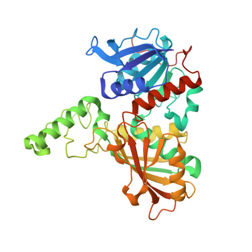
    The aspartate pathway is essential for the production of the amino acids required for protein synthesis and of the metabolites needed in bacterial development. This pathway also leads to the production of several classes of quorum-sensing molecules that can trigger virulence in certain microorganisms. The second enzyme in this pathway, aspartate β-semialdehyde dehydrogenase (ASADH), is absolutely required for bacterial survival and has been targeted for the design of selective inhibitors. Fragment-library screening has identified a new set of inhibitors that, while they do not resemble the substrates for this reaction, have been shown to bind at the active site of ASADH. Structure-guided development of these lead compounds has produced moderate inhibitors of the target enzyme, with some selectivity observed between the Gram-negative and Gram-positive orthologs of ASADH. However, many of these inhibitor analogs and derivatives have not yet achieved the expected enhanced affinity. Structural characterization of these enzyme-inhibitor complexes has provided detailed explanations for the barriers that interfere with optimal binding. Despite binding in the same active-site region, significant changes are observed in the orientation of these bound inhibitors that are caused by relatively modest structural alterations. Taken together, these studies present a cautionary tale for issues that can arise in the systematic approach to the modification of lead compounds that are being used to develop potent inhibitors.


  • Organizational Affiliation

    Department of Chemistry and Biochemistry, The University of Toledo, Toledo, OH 43606, USA.


Macromolecules
Find similar proteins by:  (by identity cutoff)  |  3D Structure
Entity ID: 1
MoleculeChains Sequence LengthOrganismDetailsImage
Aspartate-semialdehyde dehydrogenase
A, B
366Streptococcus pneumoniae SP23-BS72Mutation(s): 0 
Gene Names: asdCGSSp23BS72_03388
EC: 1.2.1.11
Entity Groups  
Sequence Clusters30% Identity50% Identity70% Identity90% Identity95% Identity100% Identity
Sequence Annotations
Expand
  • Reference Sequence
Small Molecules
Ligands 4 Unique
IDChains Name / Formula / InChI Key2D Diagram3D Interactions
NAP
Query on NAP

Download Ideal Coordinates CCD File 
D [auth A],
J [auth B],
K [auth B]
NADP NICOTINAMIDE-ADENINE-DINUCLEOTIDE PHOSPHATE
C21 H28 N7 O17 P3
XJLXINKUBYWONI-NNYOXOHSSA-N
3JL
Query on 3JL

Download Ideal Coordinates CCD File 
C [auth A],
I [auth B]
3-(3-carboxypropyl)benzene-1,2-dicarboxylic acid
C12 H12 O6
IPHJRPHIMBNGTQ-UHFFFAOYSA-N
EDO
Query on EDO

Download Ideal Coordinates CCD File 
G [auth A]
H [auth A]
L [auth B]
M [auth B]
N [auth B]
G [auth A],
H [auth A],
L [auth B],
M [auth B],
N [auth B],
O [auth B]
1,2-ETHANEDIOL
C2 H6 O2
LYCAIKOWRPUZTN-UHFFFAOYSA-N
NA
Query on NA

Download Ideal Coordinates CCD File 
E [auth A],
F [auth A]
SODIUM ION
Na
FKNQFGJONOIPTF-UHFFFAOYSA-N
Binding Affinity Annotations 
IDSourceBinding Affinity
3JL Binding MOAD:  4R4J Ki: 2.40e+6 (nM) from 1 assay(s)
Experimental Data & Validation

Experimental Data

  • Method: X-RAY DIFFRACTION
  • Resolution: 1.51 Å
  • R-Value Free: 0.190 
  • R-Value Work: 0.151 
  • R-Value Observed: 0.153 
  • Space Group: P 1 21 1
Unit Cell:
Length ( Å )Angle ( ˚ )
a = 59.925α = 90
b = 98.842β = 100.91
c = 64.329γ = 90
Software Package:
Software NamePurpose
HKL-2000data collection
REFMACrefinement
HKL-2000data reduction
SCALEPACKdata scaling

Structure Validation

View Full Validation Report


Ligand Structure Quality Assessment 


Entry History 

Deposition Data

Revision History  (Full details and data files)

  • Version 1.0: 2014-12-10
    Type: Initial release
  • Version 1.1: 2014-12-31
    Changes: Database references
  • Version 1.2: 2017-11-22
    Changes: Refinement description
  • Version 1.3: 2023-09-20
    Changes: Data collection, Database references, Derived calculations, Refinement description