Mechanism of Polyubiquitination by Human Anaphase-Promoting Complex: RING Repurposing for Ubiquitin Chain Assembly.
Brown, N.G., Watson, E.R., Weissmann, F., Jarvis, M.A., VanderLinden, R., Grace, C.R., Frye, J.J., Qiao, R., Dube, P., Petzold, G., Cho, S.E., Alsharif, O., Bao, J., Davidson, I.F., Zheng, J.J., Nourse, A., Kurinov, I., Peters, J.M., Stark, H., Schulman, B.A.(2014) Mol Cell 56: 246-260
- PubMed: 25306923 Search on PubMedSearch on PubMed Central
- DOI: https://doi.org/10.1016/j.molcel.2014.09.009
- Primary Citation Related Structures:
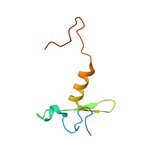
2MT5, 4R2Y - PubMed Abstract:
Polyubiquitination by E2 and E3 enzymes is a predominant mechanism regulating protein function. Some RING E3s, including anaphase-promoting complex/cyclosome (APC), catalyze polyubiquitination by sequential reactions with two different E2s. An initiating E2 ligates ubiquitin to an E3-bound substrate. Another E2 grows a polyubiquitin chain on the ubiquitin-primed substrate through poorly defined mechanisms. Here we show that human APC's RING domain is repurposed for dual functions in polyubiquitination. The canonical RING surface activates an initiating E2-ubiquitin intermediate for substrate modification. However, APC engages and activates its specialized ubiquitin chain-elongating E2 UBE2S in ways that differ from current paradigms. During chain assembly, a distinct APC11 RING surface helps deliver a substrate-linked ubiquitin to accept another ubiquitin from UBE2S. Our data define mechanisms of APC/UBE2S-mediated polyubiquitination, reveal diverse functions of RING E3s and E2s, and provide a framework for understanding distinctive RING E3 features specifying ubiquitin chain elongation.
- Department of Structural Biology, St. Jude Children's Research Hospital, Memphis, TN 38105, USA.
Organizational Affiliation: