4R2W

X-ray structure of uridine phosphorylase from Shewanella oneidensis MR-1 in complex with uridine at 1.6 A resolution


Experimental Data Snapshot

  • Method: X-RAY DIFFRACTION
  • Resolution: 1.60 Å
  • R-Value Free: 0.192 
  • R-Value Work: 0.177 
  • R-Value Observed: 0.178 

wwPDB Validation   3D Report Full Report


Ligand Structure Quality Assessment 


This is version 1.2 of the entry. See complete history


Literature

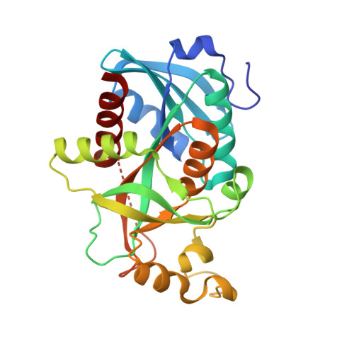
High-syn conformation of uridine and asymmetry of the hexameric molecule revealed in the high-resolution structures of Shewanella oneidensis MR-1 uridine phosphorylase in the free form and in complex with uridine.

Safonova, T.N.Mikhailov, S.N.Veiko, V.P.Mordkovich, N.N.Manuvera, V.A.Alekseev, C.S.Kovalchuk, M.V.Popov, V.O.Polyakov, K.M.

(2014) Acta Crystallogr D Biol Crystallogr 70: 3310-3319

  • DOI: https://doi.org/10.1107/S1399004714024079
  • Primary Citation of Related Structures:  
    4R2W, 4R2X

  • PubMed Abstract: 

    Uridine phosphorylase (UP; EC 2.4.2.3), a key enzyme in the pyrimidine-salvage pathway, catalyzes the reversible phosphorolysis of uridine to uracil and ribose 1-phosphate. Expression of UP from Shewanella oneidensis MR-1 (SoUP) was performed in Escherichia coli. The high-resolution X-ray structure of SoUP was solved in the free form and in complex with uridine. A crystal of SoUP in the free form was grown under microgravity and diffracted to ultrahigh resolution. Both forms of SoUP contained sulfate instead of phosphate in the active site owing to the presence of ammonium sulfate in the crystallization solution. The latter can be considered as a good mimic of phosphate. In the complex, uridine adopts a high-syn conformation with a nearly planar ribose ring and is present only in one subunit of the hexamer. A comparison of the structures of SoUP in the free form and in complex with the natural substrate uridine showed that the subunits of the hexamer are not identical, with the active sites having either an open or a closed conformation. In the monomers with the closed conformation, the active sites in which uridine is absent contain a glycerol molecule mimicking the ribose moiety of uridine.


  • Organizational Affiliation

    Bach Institute of Biochemistry, Russian Academy of Sciences, 33 Leninskii Ave., Moscow 119071, Russian Federation.


Macromolecules
Find similar proteins by:  (by identity cutoff)  |  3D Structure
Entity ID: 1
MoleculeChains Sequence LengthOrganismDetailsImage
Uridine phosphorylase
A, B, C, D, E
A, B, C, D, E, F
252Shewanella oneidensis MR-1Mutation(s): 0 
Gene Names: udpSO_4133
EC: 2.4.2.3
UniProt
Find proteins for Q8E9X9 (Shewanella oneidensis (strain MR-1))
Explore Q8E9X9 
Go to UniProtKB:  Q8E9X9
Entity Groups  
Sequence Clusters30% Identity50% Identity70% Identity90% Identity95% Identity100% Identity
UniProt GroupQ8E9X9
Sequence Annotations
Expand
  • Reference Sequence
Small Molecules
Ligands 3 Unique
IDChains Name / Formula / InChI Key2D Diagram3D Interactions
URI
Query on URI

Download Ideal Coordinates CCD File 
R [auth F]URIDINE
C9 H12 N2 O6
DRTQHJPVMGBUCF-XVFCMESISA-N
SO4
Query on SO4

Download Ideal Coordinates CCD File 
G [auth A]
H [auth A]
J [auth B]
L [auth C]
N [auth D]
G [auth A],
H [auth A],
J [auth B],
L [auth C],
N [auth D],
Q [auth E],
S [auth F],
T [auth F]
SULFATE ION
O4 S
QAOWNCQODCNURD-UHFFFAOYSA-L
GOL
Query on GOL

Download Ideal Coordinates CCD File 
I [auth A]
K [auth B]
M [auth C]
O [auth D]
P [auth D]
I [auth A],
K [auth B],
M [auth C],
O [auth D],
P [auth D],
U [auth F]
GLYCEROL
C3 H8 O3
PEDCQBHIVMGVHV-UHFFFAOYSA-N
Experimental Data & Validation

Experimental Data

  • Method: X-RAY DIFFRACTION
  • Resolution: 1.60 Å
  • R-Value Free: 0.192 
  • R-Value Work: 0.177 
  • R-Value Observed: 0.178 
  • Space Group: P 1 21 1
Unit Cell:
Length ( Å )Angle ( ˚ )
a = 91.92α = 90
b = 96.48β = 120.01
c = 91.93γ = 90
Software Package:
Software NamePurpose
HKL-2000data collection
MOLREPphasing
REFMACrefinement
HKL-2000data reduction
SCALEPACKdata scaling

Structure Validation

View Full Validation Report


Ligand Structure Quality Assessment 


Entry History 

Deposition Data

Revision History  (Full details and data files)

  • Version 1.0: 2014-12-10
    Type: Initial release
  • Version 1.1: 2015-01-07
    Changes: Database references
  • Version 1.2: 2023-09-20
    Changes: Data collection, Database references, Derived calculations, Refinement description