4R28

MspJI Restriction Endonuclease in Complex with 27-mer Oligonucleotide


Experimental Data Snapshot

  • Method: X-RAY DIFFRACTION
  • Resolution: 3.06 Å
  • R-Value Free: 0.258 
  • R-Value Work: 0.227 
  • R-Value Observed: 0.230 

wwPDB Validation   3D Report Full Report


This is version 1.2 of the entry. See complete history


Literature

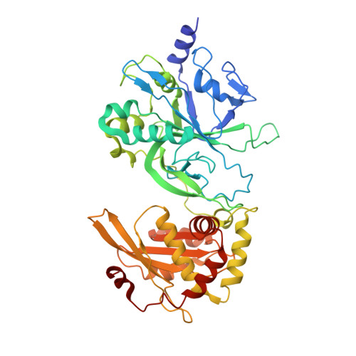
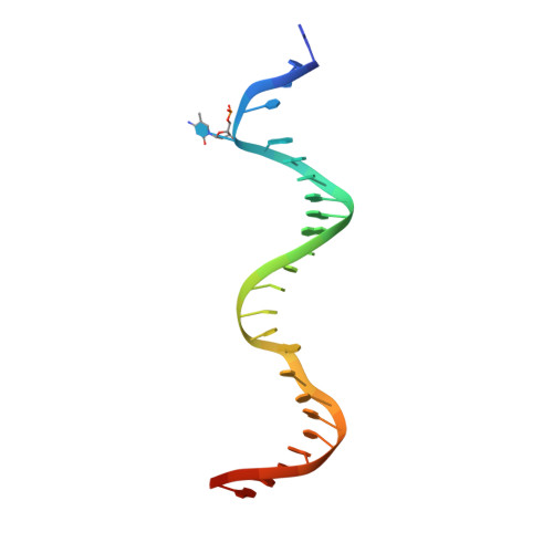
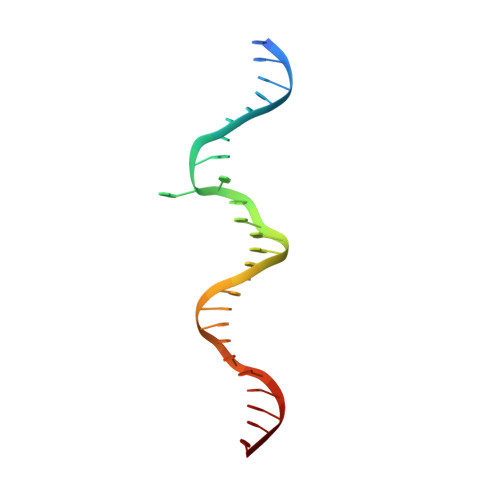
Modification-dependent restriction endonuclease, MspJI, flips 5-methylcytosine out of the DNA helix.

Horton, J.R.Wang, H.Mabuchi, M.Y.Zhang, X.Roberts, R.J.Zheng, Y.Wilson, G.G.Cheng, X.

(2014) Nucleic Acids Res 42: 12092-12101

  • DOI: https://doi.org/10.1093/nar/gku871
  • Primary Citation of Related Structures:  
    4R28

  • PubMed Abstract: 

    MspJI belongs to a family of restriction enzymes that cleave DNA containing 5-methylcytosine (5mC) or 5-hydroxymethylcytosine (5hmC). MspJI is specific for the sequence 5(h)mC-N-N-G or A and cleaves with some variability 9/13 nucleotides downstream. Earlier, we reported the crystal structure of MspJI without DNA and proposed how it might recognize this sequence and catalyze cleavage. Here we report its co-crystal structure with a 27-base pair oligonucleotide containing 5mC. This structure confirms that MspJI acts as a homotetramer and that the modified cytosine is flipped from the DNA helix into an SRA-like-binding pocket. We expected the structure to reveal two DNA molecules bound specifically to the tetramer and engaged with the enzyme's two DNA-cleavage sites. A coincidence of crystal packing precluded this organization, however. We found that each DNA molecule interacted with two adjacent tetramers, binding one specifically and the other non-specifically. The latter interaction, which prevented cleavage-site engagement, also involved base flipping and might represent the sequence-interrogation phase that precedes specific recognition. MspJI is unusual in that DNA molecules are recognized and cleaved by different subunits. Such interchange of function might explain how other complex multimeric restriction enzymes act.


  • Organizational Affiliation

    Department of Biochemistry, Emory University School of Medicine, 1510 Clifton Road, Atlanta, GA 30322, USA.


Macromolecules

Find similar proteins by:  (by identity cutoff)  |  3D Structure
Entity ID: 1
MoleculeChains Sequence LengthOrganismDetailsImage
Restriction endonuclease
A, B, C, D
456Mycobacterium sp. JLSMutation(s): 0 
Gene Names: Mjls_0822
UniProt
Find proteins for A0A0J9X157 (Mycobacterium sp. (strain JLS))
Explore A0A0J9X157 
Go to UniProtKB:  A0A0J9X157
Entity Groups  
Sequence Clusters30% Identity50% Identity70% Identity90% Identity95% Identity100% Identity
UniProt GroupA0A0J9X157
Sequence Annotations
Expand
  • Reference Sequence
Find similar nucleic acids by:  (by identity cutoff)  |  3D Structure
Entity ID: 2
MoleculeChains LengthOrganismImage
DNA (25-MER)E [auth X]27synthetic construct
Sequence Annotations
Expand
  • Reference Sequence
Find similar nucleic acids by:  (by identity cutoff)  |  3D Structure
Entity ID: 3
MoleculeChains LengthOrganismImage
DNA (26-MER)F [auth Y]27synthetic construct
Sequence Annotations
Expand
  • Reference Sequence
Experimental Data & Validation

Experimental Data

  • Method: X-RAY DIFFRACTION
  • Resolution: 3.06 Å
  • R-Value Free: 0.258 
  • R-Value Work: 0.227 
  • R-Value Observed: 0.230 
  • Space Group: P 61
Unit Cell:
Length ( Å )Angle ( ˚ )
a = 88.544α = 90
b = 88.544β = 90
c = 511.971γ = 120
Software Package:
Software NamePurpose
SERGUIdata collection
PHENIXmodel building
PHENIXrefinement
HKL-2000data reduction
HKL-2000data scaling
PHENIXphasing

Structure Validation

View Full Validation Report


Entry History 

Deposition Data

Revision History  (Full details and data files)

  • Version 1.0: 2014-10-08
    Type: Initial release
  • Version 1.1: 2014-11-19
    Changes: Database references
  • Version 1.2: 2023-09-20
    Changes: Data collection, Database references, Derived calculations, Refinement description