


Structure of a putative peptidoglycan glycosyltransferase from Atopobium parvulum in complex with cephalothin
Filippova, E.V., Minasov, G., Kiryukhina, O., Clancy, S., Joachimiak, A., Anderson, W.F.To be published.
Experimental Data Snapshot
Starting Model: experimental
View more details
Entity ID: 1 | |||||
|---|---|---|---|---|---|
| Molecule | Chains | Sequence Length | Organism | Details | Image |
| Peptidoglycan glycosyltransferase | 482 | Lancefieldella parvula DSM 20469 | Mutation(s): 0 Gene Names: Apar_1344 EC: 2.4.1.129 (PDB Primary Data), 3.4.16.4 (UniProt) | 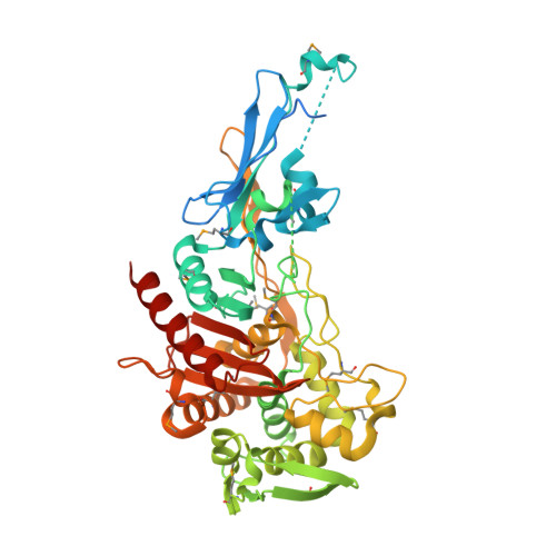 | |
UniProt | |||||
Entity Groups | |||||
| Sequence Clusters | 30% Identity50% Identity70% Identity90% Identity95% Identity100% Identity | ||||
| UniProt Group | C8W8H7 | ||||
Sequence AnnotationsExpand | |||||
Reference Sequence | |||||
| Ligands 3 Unique | |||||
|---|---|---|---|---|---|
| ID | Chains | Name / Formula / InChI Key | 2D Diagram | 3D Interactions | |
| CEP Download:Ideal Coordinates CCD File | C [auth A], F [auth B] | CEPHALOTHIN GROUP C16 H18 N2 O6 S2 UUWFGEKEQSCSMB-IAQYHMDHSA-N |  | ||
| B3P Download:Ideal Coordinates CCD File | E [auth A], H [auth B] | 2-[3-(2-HYDROXY-1,1-DIHYDROXYMETHYL-ETHYLAMINO)-PROPYLAMINO]-2-HYDROXYMETHYL-PROPANE-1,3-DIOL C11 H26 N2 O6 HHKZCCWKTZRCCL-UHFFFAOYSA-N |  | ||
| SCN Download:Ideal Coordinates CCD File | D [auth A], G [auth B] | THIOCYANATE ION C N S ZMZDMBWJUHKJPS-UHFFFAOYSA-M |  | ||
| Modified Residues 1 Unique | |||||
|---|---|---|---|---|---|
| ID | Chains | Type | Formula | 2D Diagram | Parent |
| MSE Query on MSE | A, B | L-PEPTIDE LINKING | C5 H11 N O2 Se |  | MET |
| Length ( Å ) | Angle ( ˚ ) |
|---|---|
| a = 68.553 | α = 90 |
| b = 70.002 | β = 96.76 |
| c = 115.233 | γ = 90 |
| Software Name | Purpose |
|---|---|
| Blu-Ice | data collection |
| PHASER | phasing |
| REFMAC | refinement |
| HKL-2000 | data reduction |
| HKL-2000 | data scaling |


