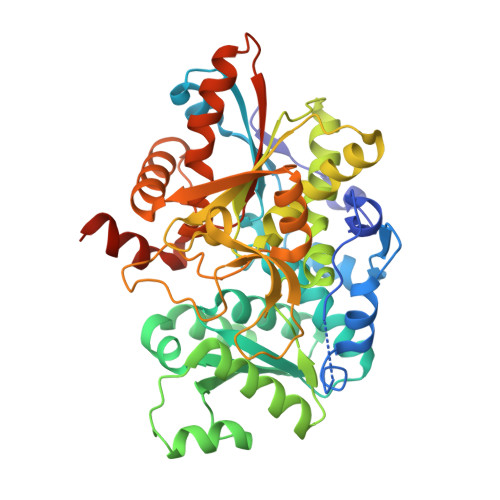
4QYS | pdb_00004qys

TrpB2 enzymes


Experimental Data Snapshot

  • Method: X-RAY DIFFRACTION
  • Resolution: 1.94 Å
  • R-Value Free: 
    0.252 (Depositor), 0.250 (DCC) 
  • R-Value Work: 
    0.195 (Depositor), 0.200 (DCC) 
  • R-Value Observed: 
    0.198 (Depositor) 

wwPDB Validation   3D Report Full Report


Ligand Structure Quality Assessment 

Created with Raphaël 2.3.0Worse 01 BetterLigand structure goodness of fit to experimental dataBest fitted PLPClick on this verticalbar to view detailsBest fitted PLRClick on this verticalbar to view detailsBest fitted SEPClick on this verticalbar to view details

This is version 1.1 of the entry. See complete history


Literature

TrpB2 enzymes are O-phospho-l-serine dependent tryptophan synthases

Busch, F.Rajendran, C.Mayans, O.Loffler, P.Merkl, R.Sterner, R.

(2014) Biochemistry 53: 6078-6083

  • DOI: https://doi.org/10.1021/bi500977y
  • Primary Citation of Related Structures:  
    4QYS

  • PubMed Abstract: 

    The rapid increase of the number of sequenced genomes asks for the functional annotation of the encoded enzymes. We used a combined computational-structural approach to determine the function of the TrpB2 subgroup of the tryptophan synthase β chain/β chain-like TrpB1-TrpB2 family (IPR023026). The results showed that TrpB2 enzymes are O-phospho-l-serine dependent tryptophan synthases, whereas TrpB1 enzymes catalyze the l-serine dependent synthesis of tryptophan. We found a single residue being responsible for the different substrate specificities of TrpB1 and TrpB2 and confirmed this finding by mutagenesis studies and crystallographic analysis of a TrpB2 enzyme with bound O-phospho-l-serine.


  • Organizational Affiliation

    Institute of Biophysics and Physical Biochemistry, University of Regensburg , Universitätsstrasse 31, D-93053 Regensburg, Germany.


Macromolecules
Find similar proteins by:  (by identity cutoff)  |  3D Structure
Entity ID: 1
MoleculeChains Sequence LengthOrganismDetailsImage
Tryptophan synthase beta chain 2A [auth B],
B [auth A]
442Saccharolobus solfataricus P2Mutation(s): 0 
Gene Names: trpB2SSO1145
EC: 4.2.1.20
UniProt
Find proteins for Q97TX6 (Saccharolobus solfataricus (strain ATCC 35092 / DSM 1617 / JCM 11322 / P2))
Explore Q97TX6 
Go to UniProtKB:  Q97TX6
Entity Groups  
Sequence Clusters30% Identity50% Identity70% Identity90% Identity95% Identity100% Identity
UniProt GroupQ97TX6
Sequence Annotations
Expand
  • Reference Sequence
Experimental Data & Validation

Experimental Data

  • Method: X-RAY DIFFRACTION
  • Resolution: 1.94 Å
  • R-Value Free:  0.252 (Depositor), 0.250 (DCC) 
  • R-Value Work:  0.195 (Depositor), 0.200 (DCC) 
  • R-Value Observed: 0.198 (Depositor) 
Space Group: P 1 21 1
Unit Cell:
Length ( Å )Angle ( ˚ )
a = 55.564α = 90
b = 61.903β = 98.23
c = 109.574γ = 90
Software Package:
Software NamePurpose
PHENIXrefinement

Structure Validation

View Full Validation Report


Ligand Structure Quality Assessment 

Created with Raphaël 2.3.0Worse 01 BetterLigand structure goodness of fit to experimental dataBest fitted PLPClick on this verticalbar to view detailsBest fitted PLRClick on this verticalbar to view detailsBest fitted SEPClick on this verticalbar to view details

Entry History 

Deposition Data

Revision History  (Full details and data files)

  • Version 1.0: 2015-02-18
    Type: Initial release
  • Version 1.1: 2024-03-20
    Changes: Data collection, Database references, Derived calculations