4QYP | pdb_00004qyp

The Crystal Structures of holo-wt human Cellular Retinol Binding protein II (hCRBPII) bound to Retinal


Experimental Data Snapshot

  • Method: X-RAY DIFFRACTION
  • Resolution: 1.62 Å
  • R-Value Free: 
    0.252 (Depositor), 0.252 (DCC) 
  • R-Value Work: 
    0.202 (Depositor), 0.203 (DCC) 
  • R-Value Observed: 
    0.204 (Depositor) 

Starting Model: experimental
View more details

wwPDB Validation 3D Report Full Report

Validation slider image for 4QYP

Ligand Structure Quality Assessment 


This is version 1.2 of the entry. See complete history

Literature

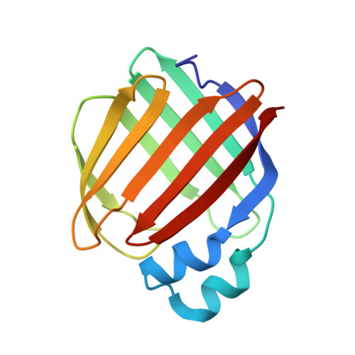
Structures of holo wild-type human cellular retinol-binding protein II (hCRBPII) bound to retinol and retinal.

Nossoni, Z.Assar, Z.Yapici, I.Nosrati, M.Wang, W.Berbasova, T.Vasileiou, C.Borhan, B.Geiger, J.

(2014) Acta Crystallogr D Biol Crystallogr 70: 3226-3232

  • DOI: https://doi.org/10.1107/S1399004714023839
  • Primary Citation Related Structures: 
    4QYN, 4QYP, 4QZT, 4QZU

  • PubMed Abstract: 

    Cellular retinol-binding proteins (CRBPs) I and II, which are members of the intracellular lipid-binding protein (iLBP) family, are retinoid chaperones that are responsible for the intracellular transport and delivery of both retinol and retinal. Although structures of retinol-bound CRBPI and CRBPII are known, no structure of a retinal-bound CRBP has been reported. In addition, the retinol-bound human CRBPII (hCRBPII) structure shows partial occupancy of a noncanonical conformation of retinol in the binding pocket. Here, the structure of retinal-bound hCRBPII and the structure of retinol-bound hCRBPII with retinol fully occupying the binding pocket are reported. It is further shown that the retinoid derivative seen in both the zebrafish CRBP and the hCRBPII structures is likely to be the product of flux-dependent and wavelength-dependent X-ray damage during data collection. The structures of retinoid-bound CRBPs are compared and contrasted, and rationales for the differences in binding affinities for retinal and retinol are provided.


  • Organizational Affiliation
    • Department of Chemistry, Michigan State University, East Lansing, MI 48864, USA.

Macromolecule Content 

  • Total Structure Weight: 63.54 kDa 
  • Atom Count: 4,761 
  • Modeled Residue Count: 532 
  • Deposited Residue Count: 532 
  • Unique protein chains: 1

Macromolecules

Find similar proteins by:|  3D Structure
Entity ID: 1
MoleculeChains  Sequence LengthOrganismDetailsImage
Retinol-binding protein 2
A, B, C, D
133Homo sapiensMutation(s): 0 
Gene Names: RBP2CRBP2
UniProt & NIH Common Fund Data Resources
Find proteins for P50120 (Homo sapiens)
Explore P50120 
Go to UniProtKB:  P50120
PHAROS:  P50120
GTEx:  ENSG00000114113 
Entity Groups
Sequence Clusters30% Identity50% Identity70% Identity90% Identity95% Identity100% Identity
UniProt GroupP50120
Sequence Annotations
Expand
Reference Sequence

Small Molecules

Ligands 2 Unique
IDChains Name / Formula / InChI Key2D Diagram3D Interactions
RET

Query on RET


Download:Ideal Coordinates CCD File
E [auth A],
F [auth B],
J [auth C]
RETINAL
C20 H28 O
NCYCYZXNIZJOKI-OVSJKPMPSA-N
ACT

Query on ACT


Download:Ideal Coordinates CCD File
G [auth B],
H [auth B],
I [auth B],
K [auth C],
L [auth D]
ACETATE ION
C2 H3 O2
QTBSBXVTEAMEQO-UHFFFAOYSA-M

Experimental Data & Validation

Experimental Data

  • Method: X-RAY DIFFRACTION
  • Resolution: 1.62 Å
  • R-Value Free:  0.252 (Depositor), 0.252 (DCC) 
  • R-Value Work:  0.202 (Depositor), 0.203 (DCC) 
  • R-Value Observed: 0.204 (Depositor) 
Space Group: P 1
Unit Cell:
Length ( Å )Angle ( ˚ )
a = 36.44α = 107.75
b = 54.892β = 97.66
c = 68.716γ = 103.08
Software Package:
Software NamePurpose
HKL-2000data collection
MOLREPphasing
REFMACrefinement
DENZOdata reduction
HKL-2000data scaling

Structure Validation

View Full Validation Report


Ligand Structure Quality Assessment 


Entry History 

Deposition Data

Revision History  (Full details and data files)

  • Version 1.0: 2014-12-10
    Type: Initial release
  • Version 1.1: 2015-01-28
    Changes: Database references
  • Version 1.2: 2023-09-20
    Changes: Data collection, Database references, Derived calculations, Refinement description