4QVI

Crystal structure of mutant ribosomal protein M218L TthL1 in complex with 80nt 23S RNA from Thermus thermophilus


Experimental Data Snapshot

  • Method: X-RAY DIFFRACTION
  • Resolution: 1.90 Å
  • R-Value Free: 0.217 
  • R-Value Work: 0.194 
  • R-Value Observed: 0.195 

wwPDB Validation   3D Report Full Report


This is version 2.1 of the entry. See complete history


Literature

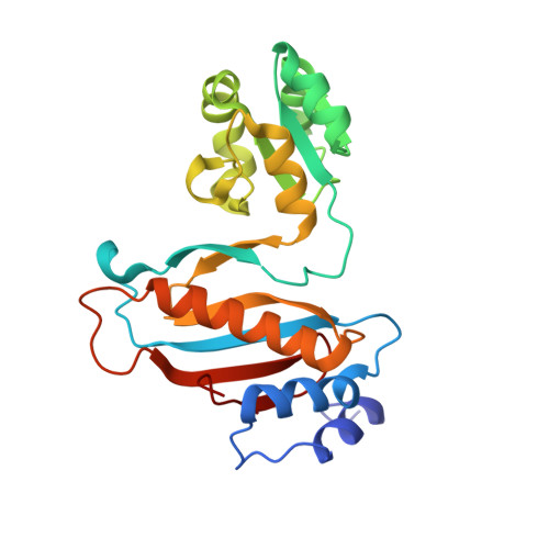
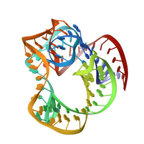
Protein-RNA affinity of ribosomal protein L1 mutants does not correlate with the number of intermolecular interactions.

Tishchenko, S.Kostareva, O.Gabdulkhakov, A.Mikhaylina, A.Nikonova, E.Nevskaya, N.Sarskikh, A.Piendl, W.Garber, M.Nikonov, S.

(2015) Acta Crystallogr D Biol Crystallogr 71: 376-386

  • DOI: https://doi.org/10.1107/S1399004714026248
  • Primary Citation of Related Structures:  
    4QG3, 4QGB, 4QVI, 4REO

  • PubMed Abstract: 

    Ribosomal protein L1, as part of the L1 stalk of the 50S ribosomal subunit, is implicated in directing tRNA movement through the ribosome during translocation. High-resolution crystal structures of four mutants (T217V, T217A, M218L and G219V) of the ribosomal protein L1 from Thermus thermophilus (TthL1) in complex with a specific 80 nt fragment of 23S rRNA and the structures of two of these mutants (T217V and G219V) in the RNA-unbound form are reported in this work. All mutations are located in the highly conserved triad Thr-Met-Gly, which is responsible for about 17% of all protein-RNA hydrogen bonds and 50% of solvent-inaccessible intermolecular hydrogen bonds. In the mutated proteins without bound RNA the RNA-binding regions show substantial conformational changes. On the other hand, in the complexes with RNA the structures of the RNA-binding surfaces in all studied mutants are very similar to the structure of the wild-type protein in complex with RNA. This shows that formation of the RNA complexes restores the distorted surfaces of the mutant proteins to a conformation characteristic of the wild-type protein complex. Domain I of the mutated TthL1 and helix 77 of 23S rRNA form a rigid body identical to that found in the complex of wild-type TthL1 with RNA, suggesting that the observed relative orientation is conserved and is probably important for ribosome function. Analysis of the complex structures and the kinetic data show that the number of intermolecular contacts and hydrogen bonds in the RNA-protein contact area does not correlate with the affinity of the protein for RNA and cannot be used as a measure of affinity.


  • Organizational Affiliation

    Institute of Protein Research, Russian Academy of Sciences, Institutskaya 4, 142290 Pushchino, Moscow Region, Russian Federation.


Macromolecules

Find similar proteins by:  (by identity cutoff)  |  3D Structure
Entity ID: 1
MoleculeChains Sequence LengthOrganismDetailsImage
50S ribosomal protein L1229Thermus thermophilusMutation(s): 1 
Gene Names: rplArpl1
UniProt
Find proteins for P27150 (Thermus thermophilus)
Explore P27150 
Go to UniProtKB:  P27150
Entity Groups  
Sequence Clusters30% Identity50% Identity70% Identity90% Identity95% Identity100% Identity
UniProt GroupP27150
Sequence Annotations
Expand
  • Reference Sequence
Find similar nucleic acids by:  (by identity cutoff)  |  3D Structure
Entity ID: 2
MoleculeChains LengthOrganismImage
fragment of 23S rRNA80Thermus thermophilus
Sequence Annotations
Expand
  • Reference Sequence
Oligosaccharides

Help

Entity ID: 3
MoleculeChains Length2D Diagram Glycosylation3D Interactions
beta-D-fructofuranose-(2-1)-alpha-D-glucopyranose
C
2N/A
Glycosylation Resources
GlyTouCan:  G05551OP
GlyCosmos:  G05551OP
Small Molecules
Ligands 3 Unique
IDChains Name / Formula / InChI Key2D Diagram3D Interactions
MES
Query on MES

Download Ideal Coordinates CCD File 
J [auth B]2-(N-MORPHOLINO)-ETHANESULFONIC ACID
C6 H13 N O4 S
SXGZJKUKBWWHRA-UHFFFAOYSA-N
SO4
Query on SO4

Download Ideal Coordinates CCD File 
D [auth A],
E [auth A],
H [auth B],
I [auth B]
SULFATE ION
O4 S
QAOWNCQODCNURD-UHFFFAOYSA-L
ACT
Query on ACT

Download Ideal Coordinates CCD File 
F [auth A],
G [auth A],
K [auth B],
L [auth B]
ACETATE ION
C2 H3 O2
QTBSBXVTEAMEQO-UHFFFAOYSA-M
Biologically Interesting Molecules (External Reference) 1 Unique
Experimental Data & Validation

Experimental Data

  • Method: X-RAY DIFFRACTION
  • Resolution: 1.90 Å
  • R-Value Free: 0.217 
  • R-Value Work: 0.194 
  • R-Value Observed: 0.195 
  • Space Group: P 21 21 21
Unit Cell:
Length ( Å )Angle ( ˚ )
a = 73.136α = 90
b = 75.845β = 90
c = 86.502γ = 90
Software Package:
Software NamePurpose
PROTEUM PLUSdata collection
PHASERphasing
PHENIXrefinement
PROTEUM PLUSdata reduction
PROTEUM PLUSdata scaling

Structure Validation

View Full Validation Report


Entry History 

Deposition Data

Revision History  (Full details and data files)

  • Version 1.0: 2015-02-11
    Type: Initial release
  • Version 1.1: 2015-02-25
    Changes: Database references
  • Version 2.0: 2020-07-29
    Type: Remediation
    Reason: Carbohydrate remediation
    Changes: Atomic model, Data collection, Database references, Derived calculations, Non-polymer description, Structure summary
  • Version 2.1: 2023-09-20
    Changes: Data collection, Database references, Refinement description, Structure summary