


Structure-based approaches towards identification of fragments for the low-druggability ATAD2 bromodomain
Chaikuad, A., Petros, A.M., Fedorov, O., Xu, J., Knapp, S.(2014) Medchemcomm 5: 1843-1848
Experimental Data Snapshot
Starting Model: experimental
View more details
(2014) Medchemcomm 5: 1843-1848
Entity ID: 1 | |||||
|---|---|---|---|---|---|
| Molecule | Chains | Sequence Length | Organism | Details | Image |
| ATPase family AAA domain-containing protein 2 | 130 | Homo sapiens | Mutation(s): 0 Gene Names: ATAD2, L16, PRO2000 EC: 3.6.1.3 (PDB Primary Data), 3.6.1 (UniProt) | 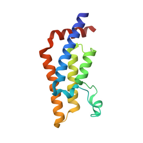 | |
UniProt & NIH Common Fund Data Resources | |||||
PHAROS: Q6PL18 GTEx: ENSG00000156802 | |||||
Entity Groups | |||||
| Sequence Clusters | 30% Identity50% Identity70% Identity90% Identity95% Identity100% Identity | ||||
| UniProt Group | Q6PL18 | ||||
Sequence AnnotationsExpand | |||||
Reference Sequence | |||||
| Ligands 3 Unique | |||||
|---|---|---|---|---|---|
| ID | Chains | Name / Formula / InChI Key | 2D Diagram | 3D Interactions | |
| 38T Download:Ideal Coordinates CCD File | H [auth A] | 5-methyluridine C10 H14 N2 O6 DWRXFEITVBNRMK-JXOAFFINSA-N |  | ||
| SO4 Download:Ideal Coordinates CCD File | B [auth A] | SULFATE ION O4 S QAOWNCQODCNURD-UHFFFAOYSA-L |  | ||
| EDO Download:Ideal Coordinates CCD File | C [auth A], D [auth A], E [auth A], F [auth A], G [auth A] | 1,2-ETHANEDIOL C2 H6 O2 LYCAIKOWRPUZTN-UHFFFAOYSA-N |  | ||
| Length ( Å ) | Angle ( ˚ ) |
|---|---|
| a = 79.59 | α = 90 |
| b = 79.59 | β = 90 |
| c = 138.421 | γ = 120 |
| Software Name | Purpose |
|---|---|
| CrystalClear | data collection |
| PHASER | phasing |
| REFMAC | refinement |
| MOSFLM | data reduction |
| SCALA | data scaling |


