


Structural characterization of potential modulation sites in the extracellular domain of the prokaryotic pentameric proton-gated ion channel GLIC
Fourati, Z., Sauguet, L., Delarue, M.(2015) Acta Crystallogr D Biol Crystallogr
Experimental Data Snapshot
(2015) Acta Crystallogr D Biol Crystallogr
Entity ID: 1 | |||||
|---|---|---|---|---|---|
| Molecule | Chains | Sequence Length | Organism | Details | Image |
| Proton-gated ion channel | 316 | Gloeobacter violaceus PCC 7421 | Mutation(s): 0 Gene Names: glvI, glr4197 Membrane Entity: Yes | 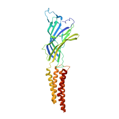 | |
UniProt | |||||
Entity Groups | |||||
| Sequence Clusters | 30% Identity50% Identity70% Identity90% Identity95% Identity100% Identity | ||||
| UniProt Group | Q7NDN8 | ||||
Sequence AnnotationsExpand | |||||
Reference Sequence | |||||
| Ligands 4 Unique | |||||
|---|---|---|---|---|---|
| ID | Chains | Name / Formula / InChI Key | 2D Diagram | 3D Interactions | |
| PLC Download:Ideal Coordinates CCD File | P [auth C], V [auth E] | DIUNDECYL PHOSPHATIDYL CHOLINE C32 H65 N O8 P IJFVSSZAOYLHEE-SSEXGKCCSA-O |  | ||
| LMT Download:Ideal Coordinates CCD File | F [auth A] G [auth A] I [auth B] M [auth C] Q [auth D] | DODECYL-BETA-D-MALTOSIDE C24 H46 O11 NLEBIOOXCVAHBD-QKMCSOCLSA-N |  | ||
| CL Download:Ideal Coordinates CCD File | H [auth A], J [auth B], N [auth C], S [auth D], U [auth E] | CHLORIDE ION Cl VEXZGXHMUGYJMC-UHFFFAOYSA-M |  | ||
| NA Download:Ideal Coordinates CCD File | K [auth B], L [auth C], O [auth C], R [auth D] | SODIUM ION Na FKNQFGJONOIPTF-UHFFFAOYSA-N |  | ||
| Length ( Å ) | Angle ( ˚ ) |
|---|---|
| a = 181.878 | α = 90 |
| b = 134.423 | β = 102.72 |
| c = 160.003 | γ = 90 |
| Software Name | Purpose |
|---|---|
| XDS | data scaling |
| REFMAC | refinement |
| BUSTER | refinement |
| XDS | data reduction |
| SCALA | data scaling |
| REFMAC | phasing |


