Structures and binding studies of the complexes of phospholipase A2 with five inhibitors
Shukla, P.K., Gautam, L., Sinha, M., Kaur, P., Sharma, S., Singh, T.P.(2015) Biochim Biophys Acta 1854: 269-277
- PubMed: 25541253
- DOI: https://doi.org/10.1016/j.bbapap.2014.12.017
- Primary Citation of Related Structures:
4QEM, 4QER, 4QF7, 4QF8, 4QGD - PubMed Abstract:
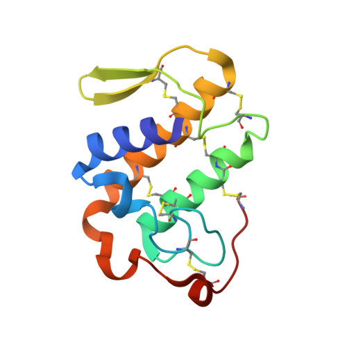
Phospholipase A2 (PLA2) catalyzes the hydrolysis of phospholipids into arachidonic acid and lysophospholipids. Arachidonic acid is used as a substrate in the next step of the multistep pathway leading to the production of eicosanoids. The eicosanoids, in extremely low concentrations, are required in a number of physiological processes. However, the increase in their concentrations above the essential physiological requirements leads to various inflammatory conditions. In order to prevent the unwanted rise in the concentrations of eicosanoids, the actions of PLA2 and other enzymes of the pathway need to be blocked. We report here the structures of five complexes of group IIA PLA2 from Daboia russelli pulchella with tightly binding inhibitors, (i) p-coumaric acid, (ii) resveratrol, (iii) spermidine, (iv) corticosterone and (v) gramine derivative. The binding studies using fluorescence spectroscopy and surface plasmon resonance techniques for the interactions of PLA2 with the above five compounds showed high binding affinities with values of dissociation constants (KD) ranging from 3.7×10(-8) M to 2.1×10(-9) M. The structure determinations of the complexes of PLA2 with the above five compounds showed that all the compounds bound to PLA2 in the substrate binding cleft. The protein residues that contributed to the interactions with these compounds included Leu2, Leu3, Phe5, Gly6, Ile9, Ala18, Ile19, Trp22, Ser23, Cys29, Gly30, Cys45, His48, Asp49 and Phe106. The positions of side chains of several residues including Leu2, Leu3, Ile19, Trp31, Lys69, Ser70 and Arg72 got significantly shifted while the positions of active site residues, His48, Asp49, Tyr52 and Asp99 were unperturbed.
- Department of Biophysics, All India Institute of Medical Sciences, New Delhi, India.
Organizational Affiliation:


