4Q1U

Serum paraoxonase-1 by directed evolution with the K192Q mutation


Experimental Data Snapshot

  • Method: X-RAY DIFFRACTION
  • Resolution: 2.30 Å
  • R-Value Free: 0.212 
  • R-Value Work: 0.176 
  • R-Value Observed: 0.178 

wwPDB Validation   3D Report Full Report


Ligand Structure Quality Assessment 


This is version 1.2 of the entry. See complete history


Literature

Catalytic stimulation by restrained active-site floppiness-the case of high density lipoprotein-bound serum paraoxonase-1.

Ben-David, M.Sussman, J.L.Maxwell, C.I.Szeler, K.Kamerlin, S.C.Tawfik, D.S.

(2015) J Mol Biol 427: 1359-1374

  • DOI: https://doi.org/10.1016/j.jmb.2015.01.013
  • Primary Citation of Related Structures:  
    4Q1U

  • PubMed Abstract: 

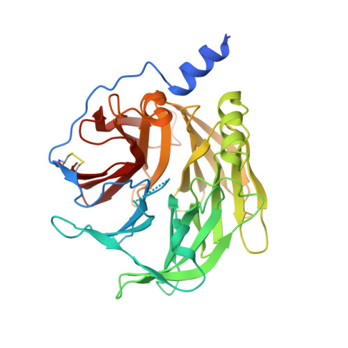
    Despite the abundance of membrane-associated enzymes, the mechanism by which membrane binding stabilizes these enzymes and stimulates their catalysis remains largely unknown. Serum paraoxonase-1 (PON1) is a lipophilic lactonase whose stability and enzymatic activity are dramatically stimulated when associated with high-density lipoprotein (HDL) particles. Our mutational and structural analyses, combined with empirical valence bond simulations, reveal a network of hydrogen bonds that connect HDL binding residues with Asn168--a key catalytic residue residing >15Å from the HDL contacting interface. This network ensures precise alignment of N168, which, in turn, ligates PON1's catalytic calcium and aligns the lactone substrate for catalysis. HDL binding restrains the overall motion of the active site and particularly of N168, thus reducing the catalytic activation energy barrier. We demonstrate herein that disturbance of this network, even at its most far-reaching periphery, undermines PON1's activity. Membrane binding thus immobilizes long-range interactions via second- and third-shell residues that reduce the active site's floppiness and pre-organize the catalytic residues. Although this network is critical for efficient catalysis, as demonstrated here, unraveling these long-rage interaction networks is challenging, let alone their implementation in artificial enzyme design.


  • Organizational Affiliation

    Department of Structural Biology, Weizmann Institute of Science, Rehovot 76100, Israel; Department of Biological Chemistry, Weizmann Institute of Science, Rehovot 76100, Israel.


Macromolecules
Find similar proteins by:  (by identity cutoff)  |  3D Structure
Entity ID: 1
MoleculeChains Sequence LengthOrganismDetailsImage
Serum paraoxonase/arylesterase 1355hybridMutation(s): 0 
EC: 3.1.1.2 (PDB Primary Data), 3.1.1.81 (PDB Primary Data), 3.1.8.1 (PDB Primary Data)
Entity Groups  
Sequence Clusters30% Identity50% Identity70% Identity90% Identity95% Identity100% Identity
Sequence Annotations
Expand
  • Reference Sequence
Small Molecules
Ligands 5 Unique
IDChains Name / Formula / InChI Key2D Diagram3D Interactions
LMT
Query on LMT

Download Ideal Coordinates CCD File 
D [auth A]DODECYL-BETA-D-MALTOSIDE
C24 H46 O11
NLEBIOOXCVAHBD-QKMCSOCLSA-N
PO4
Query on PO4

Download Ideal Coordinates CCD File 
E [auth A]PHOSPHATE ION
O4 P
NBIIXXVUZAFLBC-UHFFFAOYSA-K
BR
Query on BR

Download Ideal Coordinates CCD File 
G [auth A]BROMIDE ION
Br
CPELXLSAUQHCOX-UHFFFAOYSA-M
CA
Query on CA

Download Ideal Coordinates CCD File 
B [auth A],
C [auth A]
CALCIUM ION
Ca
BHPQYMZQTOCNFJ-UHFFFAOYSA-N
CL
Query on CL

Download Ideal Coordinates CCD File 
F [auth A]CHLORIDE ION
Cl
VEXZGXHMUGYJMC-UHFFFAOYSA-M
Experimental Data & Validation

Experimental Data

  • Method: X-RAY DIFFRACTION
  • Resolution: 2.30 Å
  • R-Value Free: 0.212 
  • R-Value Work: 0.176 
  • R-Value Observed: 0.178 
  • Space Group: P 43 21 2
Unit Cell:
Length ( Å )Angle ( ˚ )
a = 93.514α = 90
b = 93.514β = 90
c = 144.007γ = 90
Software Package:
Software NamePurpose
HKL-2000data collection
PHASERphasing
REFMACrefinement
HKL-2000data reduction
HKL-2000data scaling

Structure Validation

View Full Validation Report


Ligand Structure Quality Assessment 


Entry History 

Deposition Data

Revision History  (Full details and data files)

  • Version 1.0: 2015-02-18
    Type: Initial release
  • Version 1.1: 2015-03-25
    Changes: Database references
  • Version 1.2: 2023-09-20
    Changes: Data collection, Database references, Derived calculations, Refinement description