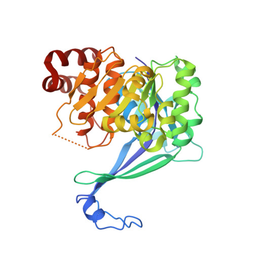
4PVV

Micobacterial Adenosine Kinase in complex with inhibitor


Experimental Data Snapshot

  • Method: X-RAY DIFFRACTION
  • Resolution: 2.50 Å
  • R-Value Free: 0.341 
  • R-Value Work: 0.264 
  • R-Value Observed: 0.268 

wwPDB Validation   3D Report Full Report


Ligand Structure Quality Assessment 


This is version 1.1 of the entry. See complete history


Literature

Structural Basis for Inhibition of Mycobacterial and Human Adenosine Kinase by 7-Substituted 7-(Het)aryl-7-deazaadenine Ribonucleosides

Snasel, J.Naus, P.Dostal, J.Hnizda, A.Fanfrlik, J.Brynda, J.Bourderioux, A.Dusek, M.Dvorakova, H.Stolarikova, J.Zabranska, H.Pohl, R.Konecny, P.Dzubak, P.Votruba, I.Hajduch, M.Rezacova, P.Veverka, V.Hocek, M.Pichova, I.

(2014) J Med Chem 57: 8268-8279

  • DOI: https://doi.org/10.1021/jm500497v
  • Primary Citation of Related Structures:  
    4O1G, 4O1L, 4PVV

  • PubMed Abstract: 

    Adenosine kinase (ADK) from Mycobacterium tuberculosis (Mtb) was selected as a target for design of antimycobacterial nucleosides. Screening of 7-(het)aryl-7-deazaadenine ribonucleosides with Mtb and human (h) ADKs and testing with wild-type and drug-resistant Mtb strains identified specific inhibitors of Mtb ADK with micromolar antimycobacterial activity and low cytotoxicity. X-ray structures of complexes of Mtb and hADKs with 7-ethynyl-7-deazaadenosine showed differences in inhibitor interactions in the adenosine binding sites. 1D (1)H STD NMR experiments revealed that these inhibitors are readily accommodated into the ATP and adenosine binding sites of Mtb ADK, whereas they bind preferentially into the adenosine site of hADK. Occupation of the Mtb ADK ATP site with inhibitors and formation of catalytically less competent semiopen conformation of MtbADK after inhibitor binding in the adenosine site explain the lack of phosphorylation of 7-substituted-7-deazaadenosines. Semiempirical quantum mechanical analysis confirmed different affinity of nucleosides for the Mtb ADK adenosine and ATP sites.


  • Organizational Affiliation

    Institute of Organic Chemistry and Biochemistry, Academy of Sciences of the Czech Republic , Flemingovo nam. 2, 16610 Prague 6, Czech Republic.


Macromolecules
Find similar proteins by:  (by identity cutoff)  |  3D Structure
Entity ID: 1
MoleculeChains Sequence LengthOrganismDetailsImage
Adenosine kinase324Mycobacterium tuberculosisMutation(s): 0 
Gene Names: adoK
EC: 2.7.1.20
UniProt
Find proteins for P9WID5 (Mycobacterium tuberculosis (strain ATCC 25618 / H37Rv))
Explore P9WID5 
Go to UniProtKB:  P9WID5
Entity Groups  
Sequence Clusters30% Identity50% Identity70% Identity90% Identity95% Identity100% Identity
UniProt GroupP9WID5
Sequence Annotations
Expand
  • Reference Sequence
Small Molecules
Ligands 1 Unique
IDChains Name / Formula / InChI Key2D Diagram3D Interactions
HO4
Query on HO4

Download Ideal Coordinates CCD File 
B [auth A]5-ethynyl-7-(beta-D-ribofuranosyl)-7H-pyrrolo[2,3-d]pyrimidin-4-amine
C13 H14 N4 O4
ANCWCJFYCNNXDR-QYVSTXNMSA-N
Binding Affinity Annotations 
IDSourceBinding Affinity
HO4 Binding MOAD:  4PVV IC50: 330 (nM) from 1 assay(s)
Experimental Data & Validation

Experimental Data

  • Method: X-RAY DIFFRACTION
  • Resolution: 2.50 Å
  • R-Value Free: 0.341 
  • R-Value Work: 0.264 
  • R-Value Observed: 0.268 
  • Space Group: P 31 1 2
Unit Cell:
Length ( Å )Angle ( ˚ )
a = 46.985α = 90
b = 46.985β = 90
c = 244.25γ = 120
Software Package:
Software NamePurpose
MOSFLMdata reduction
SCALAdata scaling
MOLREPphasing
REFMACrefinement
PDB_EXTRACTdata extraction

Structure Validation

View Full Validation Report


Ligand Structure Quality Assessment 


Entry History 

Deposition Data

Revision History  (Full details and data files)

  • Version 1.0: 2014-11-26
    Type: Initial release
  • Version 1.1: 2024-03-20
    Changes: Data collection, Database references, Derived calculations