A loose domain swapping organization confers a remarkable stability to the dimeric structure of the arginine binding protein from Thermotoga maritima
Ruggiero, A., Dattelbaum, J.D., Staiano, M., Berisio, R., D'Auria, S., Vitagliano, L.(2014) PLoS One 9: e96560-e96560
- PubMed: 24832102 Search on PubMedSearch on PubMed Central
- DOI: https://doi.org/10.1371/journal.pone.0096560
- Primary Citation Related Structures:
4PRS, 4PSH - PubMed Abstract:
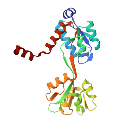
The arginine binding protein from Thermatoga maritima (TmArgBP), a substrate binding protein (SBP) involved in the ABC system of solute transport, presents a number of remarkable properties. These include an extraordinary stability to temperature and chemical denaturants and the tendency to form multimeric structures, an uncommon feature among SBPs involved in solute transport. Here we report a biophysical and structural characterization of the TmArgBP dimer. Our data indicate that the dimer of the protein is endowed with a remarkable stability since its full dissociation requires high temperature as well as SDS and urea at high concentrations. In order to elucidate the atomic level structural properties of this intriguing protein, we determined the crystallographic structures of the apo and the arginine-bound forms of TmArgBP using MAD and SAD methods, respectively. The comparison of the liganded and unliganded models demonstrates that TmArgBP tertiary structure undergoes a very large structural re-organization upon arginine binding. This transition follows the Venus Fly-trap mechanism, although the entity of the re-organization observed in TmArgBP is larger than that observed in homologous proteins. Intriguingly, TmArgBP dimerizes through the swapping of the C-terminal helix. This dimer is stabilized exclusively by the interactions established by the swapping helix. Therefore, the TmArgBP dimer combines a high level of stability and conformational freedom. The structure of the TmArgBP dimer represents an uncommon example of large tertiary structure variations amplified at quaternary structure level by domain swapping. Although the biological relevance of the dimer needs further assessments, molecular modelling suggests that the two TmArgBP subunits may simultaneously interact with two distinct ABC transporters. Moreover, the present protein structures provide some clues about the determinants of the extraordinary stability of the biomolecule. The availability of an accurate 3D model represents a powerful tool for the design of new TmArgBP suited for biotechnological applications.
- Institute of Biostructures and Bioimaging, CNR, Napoli, Italy.
Organizational Affiliation: