


Crystal structure of a phosphate binding protein
Opatowsky, Y., Neznansky, A.To be published.
Experimental Data Snapshot
wwPDB Validation 3D Report Full Report

Entity ID: 1 | |||||
|---|---|---|---|---|---|
| Molecule | Chains | Sequence Length | Organism | Details | Image |
| Phosphate binding protein | A, B [auth C] | 299 | Pseudomonas aeruginosa | Mutation(s): 0 Gene Names: pstS | 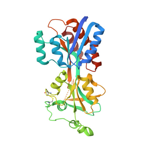 |
UniProt | |||||
Entity Groups | |||||
| Sequence Clusters | 30% Identity50% Identity70% Identity90% Identity95% Identity100% Identity | ||||
| UniProt Group | P0DMR4 | ||||
Sequence AnnotationsExpand | |||||
Reference Sequence | |||||
| Ligands 1 Unique | |||||
|---|---|---|---|---|---|
| ID | Chains | Name / Formula / InChI Key | 2D Diagram | 3D Interactions | |
| PO4 Download:Ideal Coordinates CCD File | C [auth A], D [auth C] | PHOSPHATE ION O4 P NBIIXXVUZAFLBC-UHFFFAOYSA-K |  | ||
| Length ( Å ) | Angle ( ˚ ) |
|---|---|
| a = 67.5 | α = 90 |
| b = 151.35 | β = 90 |
| c = 108.98 | γ = 90 |
| Software Name | Purpose |
|---|---|
| EDNA | data collection |
| PHASER | phasing |
| PHENIX | refinement |
| MOSFLM | data reduction |
| SCALA | data scaling |


