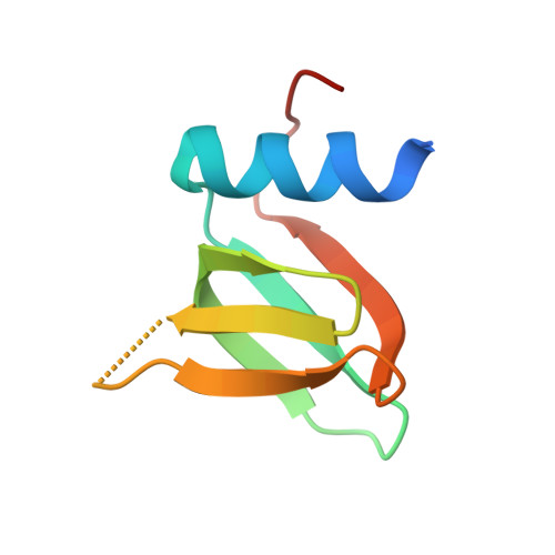
4PNO

Escherichia coli Hfq-RNA complex at 0.97 A Resolution


Experimental Data Snapshot

  • Method: X-RAY DIFFRACTION
  • Resolution: 0.97 Å
  • R-Value Free: 0.164 
  • R-Value Work: 0.142 
  • R-Value Observed: 0.144 

Starting Model: experimental
View more details

wwPDB Validation   3D Report Full Report


Ligand Structure Quality Assessment 


This is version 1.3 of the entry. See complete history


Literature

Structure of an Escherichia coli Hfq:RNA complex at 0.97 angstrom resolution.

Schulz, E.C.Barabas, O.

(2014) Acta Crystallogr F Struct Biol Commun 70: 1492-1497

  • DOI: https://doi.org/10.1107/S2053230X14020044
  • Primary Citation of Related Structures:  
    4PNO

  • PubMed Abstract: 

    In bacteria, small RNAs (sRNAs) silence or activate target genes through base pairing with the mRNA, thereby modulating its translation. A central player in this process is the RNA chaperone Hfq, which facilitates the annealing of sRNAs with their target mRNAs. Hfq has two RNA-binding surfaces that recognize A-rich and U-rich sequences, and is believed to bind an sRNA-mRNA pair simultaneously. However, how Hfq promotes annealing remains unclear. Here, the crystal structure of Escherichia coli Hfq is presented in complex with U6-RNA bound to its proximal binding site at 0.97 Å resolution, revealing the Hfq-RNA interaction in exceptional detail.


  • Organizational Affiliation

    Structural and Computational Biology Unit, European Molecular Biology Laboratory, Meyerhofstrasse 1, 69117 Heidelberg, Germany.


Macromolecules
Find similar proteins by:  (by identity cutoff)  |  3D Structure
Entity ID: 1
MoleculeChains Sequence LengthOrganismDetailsImage
RNA-binding protein Hfq72Escherichia coliMutation(s): 0 
Gene Names: hfqBN17_41491BU34_16545ECs5148LF82_0993
UniProt
Find proteins for C1IFD2 (Escherichia coli)
Explore C1IFD2 
Go to UniProtKB:  C1IFD2
Entity Groups  
Sequence Clusters30% Identity50% Identity70% Identity90% Identity95% Identity100% Identity
UniProt GroupC1IFD2
Sequence Annotations
Expand
  • Reference Sequence
Small Molecules
Ligands 1 Unique
IDChains Name / Formula / InChI Key2D Diagram3D Interactions
U5P
Query on U5P

Download Ideal Coordinates CCD File 
B [auth A]URIDINE-5'-MONOPHOSPHATE
C9 H13 N2 O9 P
DJJCXFVJDGTHFX-XVFCMESISA-N
Experimental Data & Validation

Experimental Data

  • Method: X-RAY DIFFRACTION
  • Resolution: 0.97 Å
  • R-Value Free: 0.164 
  • R-Value Work: 0.142 
  • R-Value Observed: 0.144 
  • Space Group: P 6
Unit Cell:
Length ( Å )Angle ( ˚ )
a = 61.11α = 90
b = 61.11β = 90
c = 27.86γ = 120
Software Package:
Software NamePurpose
PHENIXrefinement

Structure Validation

View Full Validation Report


Ligand Structure Quality Assessment 


Entry History & Funding Information

Deposition Data


Funding OrganizationLocationGrant Number
EU FP7 - Marie Curie IEFGermanyPIEF-GA-2011-301489

Revision History  (Full details and data files)

  • Version 1.0: 2014-11-19
    Type: Initial release
  • Version 1.1: 2014-11-26
    Changes: Data collection
  • Version 1.2: 2018-03-07
    Changes: Advisory, Data collection, Derived calculations, Other, Source and taxonomy
  • Version 1.3: 2023-09-27
    Changes: Advisory, Data collection, Database references, Refinement description