4PKL | pdb_00004pkl

Bromodomain of Trypanosoma brucei BDF2 With IBET-151


Experimental Data Snapshot

  • Method: X-RAY DIFFRACTION
  • Resolution: 1.25 Å
  • R-Value Free: 
    0.179 (Depositor), 0.181 (DCC) 
  • R-Value Work: 
    0.169 (Depositor), 0.172 (DCC) 
  • R-Value Observed: 
    0.170 (Depositor) 

wwPDB Validation 3D Report Full Report

Validation slider image for 4PKL

Ligand Structure Quality Assessment 


This is version 1.3 of the entry. See complete history

Literature

Bromodomain Proteins Contribute to Maintenance of Bloodstream Form Stage Identity in the African Trypanosome.

Schulz, D.Mugnier, M.R.Paulsen, E.M.Kim, H.S.Chung, C.W.Tough, D.F.Rioja, I.Prinjha, R.K.Papavasiliou, F.N.Debler, E.W.

(2015) PLoS Biol 13: e1002316-e1002316

  • DOI: https://doi.org/10.1371/journal.pbio.1002316
  • Primary Citation Related Structures: 
    4PKL

  • PubMed Abstract: 

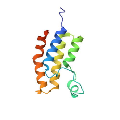
    Trypanosoma brucei, the causative agent of African sleeping sickness, is transmitted to its mammalian host by the tsetse. In the fly, the parasite's surface is covered with invariant procyclin, while in the mammal it resides extracellularly in its bloodstream form (BF) and is densely covered with highly immunogenic Variant Surface Glycoprotein (VSG). In the BF, the parasite varies this highly immunogenic surface VSG using a repertoire of ~2500 distinct VSG genes. Recent reports in mammalian systems point to a role for histone acetyl-lysine recognizing bromodomain proteins in the maintenance of stem cell fate, leading us to hypothesize that bromodomain proteins may maintain the BF cell fate in trypanosomes. Using small-molecule inhibitors and genetic mutants for individual bromodomain proteins, we performed RNA-seq experiments that revealed changes in the transcriptome similar to those seen in cells differentiating from the BF to the insect stage. This was recapitulated at the protein level by the appearance of insect-stage proteins on the cell surface. Furthermore, bromodomain inhibition disrupts two major BF-specific immune evasion mechanisms that trypanosomes harness to evade mammalian host antibody responses. First, monoallelic expression of the antigenically varied VSG is disrupted. Second, rapid internalization of antibodies bound to VSG on the surface of the trypanosome is blocked. Thus, our studies reveal a role for trypanosome bromodomain proteins in maintaining bloodstream stage identity and immune evasion. Importantly, bromodomain inhibition leads to a decrease in virulence in a mouse model of infection, establishing these proteins as potential therapeutic drug targets for trypanosomiasis. Our 1.25Å resolution crystal structure of a trypanosome bromodomain in complex with I-BET151 reveals a novel binding mode of the inhibitor, which serves as a promising starting point for rational drug design.


  • Organizational Affiliation
    • Laboratory of Lymphocyte Biology, The Rockefeller University, New York, New York, United States of America.

Macromolecule Content 

  • Total Structure Weight: 28.13 kDa 
  • Atom Count: 2,113 
  • Modeled Residue Count: 218 
  • Deposited Residue Count: 238 
  • Unique protein chains: 1

Macromolecules

Find similar proteins by:|  3D Structure
Entity ID: 1
MoleculeChains  Sequence LengthOrganismDetailsImage
Bromodomain factor 2
A, B
119Trypanosoma brucei brucei TREU927Mutation(s): 0 
Gene Names: Tb10.6k15.3240
UniProt
Find proteins for Q38AM1 (Trypanosoma brucei brucei (strain 927/4 GUTat10.1))
Explore Q38AM1 
Go to UniProtKB:  Q38AM1
Entity Groups
Sequence Clusters30% Identity50% Identity70% Identity90% Identity95% Identity100% Identity
UniProt GroupQ38AM1
Sequence Annotations
Expand
Reference Sequence

Small Molecules

Ligands 5 Unique
IDChains Name / Formula / InChI Key2D Diagram3D Interactions
1GH

Query on 1GH


Download:Ideal Coordinates CCD File
C [auth A],
G [auth B]
7-(3,5-DIMETHYL-1,2-OXAZOL-4-YL)-8-METHOXY-1-[(1R)-1-(PYRIDIN-2-YL)ETHYL]-1H,2H,3H-IMIDAZO[4,5-C]QUINOLIN-2-ONE
C23 H21 N5 O3
VUVUVNZRUGEAHB-CYBMUJFWSA-N
PO4

Query on PO4


Download:Ideal Coordinates CCD File
D [auth A]PHOSPHATE ION
O4 P
NBIIXXVUZAFLBC-UHFFFAOYSA-K
GOL

Query on GOL


Download:Ideal Coordinates CCD File
I [auth B]GLYCEROL
C3 H8 O3
PEDCQBHIVMGVHV-UHFFFAOYSA-N
K

Query on K


Download:Ideal Coordinates CCD File
E [auth A],
H [auth B]
POTASSIUM ION
K
NPYPAHLBTDXSSS-UHFFFAOYSA-N
CL

Query on CL


Download:Ideal Coordinates CCD File
F [auth A]CHLORIDE ION
Cl
VEXZGXHMUGYJMC-UHFFFAOYSA-M

Experimental Data & Validation

Experimental Data

  • Method: X-RAY DIFFRACTION
  • Resolution: 1.25 Å
  • R-Value Free:  0.179 (Depositor), 0.181 (DCC) 
  • R-Value Work:  0.169 (Depositor), 0.172 (DCC) 
  • R-Value Observed: 0.170 (Depositor) 
Space Group: P 21 21 21
Unit Cell:
Length ( Å )Angle ( ˚ )
a = 55.869α = 90
b = 62.091β = 90
c = 62.117γ = 90
Software Package:
Software NamePurpose
PHENIXrefinement
ARPmodel building
HKL-2000data reduction
PHASERphasing
Cootmodel building
HKL-2000data scaling
Omodel building

Structure Validation

View Full Validation Report


Ligand Structure Quality Assessment 


Entry History 

& Funding Information

Deposition Data

  • Released Date: 2015-11-18 
  • Deposition Author(s): Debler, E.W.

Funding OrganizationLocationGrant Number
Bill & Melinda Gates FoundationUnited StatesOPP1108062

Revision History  (Full details and data files)

  • Version 1.0: 2015-11-18
    Type: Initial release
  • Version 1.1: 2017-09-27
    Changes: Author supporting evidence, Derived calculations
  • Version 1.2: 2020-09-16
    Changes: Database references, Derived calculations
  • Version 1.3: 2023-12-27
    Changes: Data collection, Database references