Characterization of Variants of the Pore-Forming Toxin ClyA from Escherichia coli Controlled by a Redox Switch.
Roderer, D., Benke, S., Muller, M., Fah-Rechsteiner, H., Ban, N., Schuler, B., Glockshuber, R.(2014) Biochemistry 53: 6357-6369
- PubMed: 25222267 Search on PubMed
- DOI: https://doi.org/10.1021/bi5007578
- Primary Citation Related Structures:
4PHO, 4PHQ - PubMed Abstract:
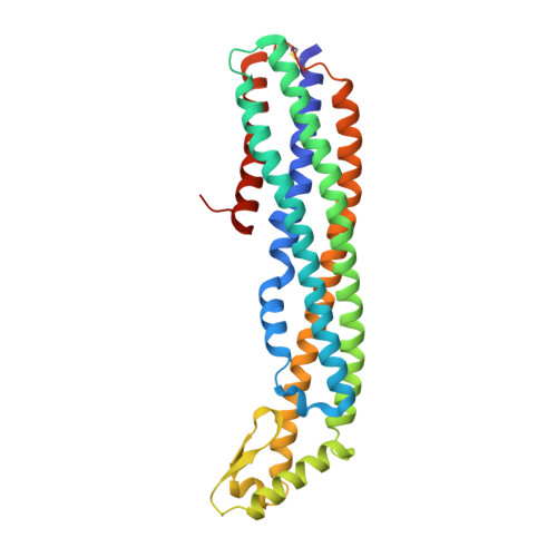
The α-pore-forming toxin Cytolysin A (ClyA) is responsible for the hemolytic phenotype of several Escherichia coli and Salmonella enterica strains. ClyA is a soluble, 34 kDa monomer that assembles into a dodecameric pore complex in the presence of membranes or detergent. The comparison of the X-ray structures of monomeric ClyA and the ClyA protomer in the pore complex revealed one of the largest conformational transitions observed so far in proteins, involving the structural rearrangement of more than half of all residues, which is consistent with the finding that conversion from the monomer to the assembly competent protomer is rate-limiting for pore assembly. Here, we introduced artificial disulfide bonds at two distinct sites into the ClyA monomer that both prevent a specific structural rearrangement required for protomer formation. Using electron microscopy and hemolytic activity assays, we show that the engineered disulfides indeed trap these ClyA variants in an assembly incompetent state. Assembly of the variants into functional pore complexes can be completely recovered by disulfide reduction. The assembly kinetics of the ClyA variants recorded with circular dichroism and fluorescence spectroscopy revealed the same mechanism of protomer formation that was observed for wild-type ClyA, proceeding via an intermediate with decreased secondary structure content.
- Institute of Molecular Biology and Biophysics, ETH Zürich , Otto-Stern-Weg 5, CH-8093 Zürich, Switzerland.
Organizational Affiliation: