INTRACELLULAR TRANSPORT. Phosphatidylserine transport by ORP/Osh proteins is driven by phosphatidylinositol 4-phosphate.
Moser von Filseck, J., Copic, A., Delfosse, V., Vanni, S., Jackson, C.L., Bourguet, W., Drin, G.(2015) Science 349: 432-436
- PubMed: 26206936 Search on PubMed
- DOI: https://doi.org/10.1126/science.aab1346
- Primary Citation Related Structures:
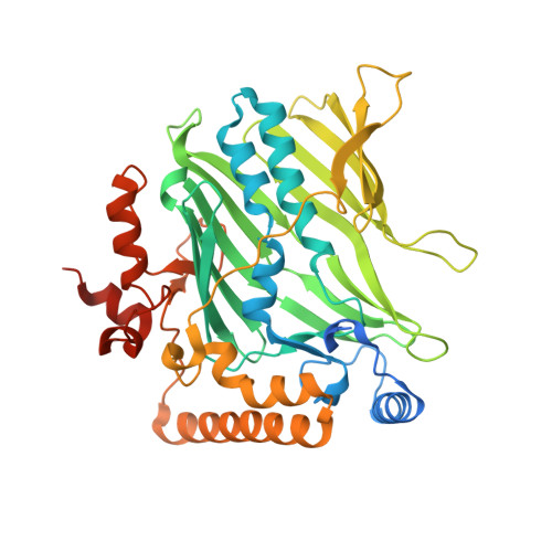
4PH7 - PubMed Abstract:
In eukaryotic cells, phosphatidylserine (PS) is synthesized in the endoplasmic reticulum (ER) but is highly enriched in the plasma membrane (PM), where it contributes negative charge and to specific recruitment of signaling proteins. This distribution relies on transport mechanisms whose nature remains elusive. Here, we found that the PS transporter Osh6p extracted phosphatidylinositol 4-phosphate (PI4P) and exchanged PS for PI4P between two membranes. We solved the crystal structure of Osh6p:PI4P complex and demonstrated that the transport of PS by Osh6p depends on PI4P recognition in vivo. Finally, we showed that the PI4P-phosphatase Sac1p, by maintaining a PI4P gradient at the ER/PM interface, drove PS transport. Thus, PS transport by oxysterol-binding protein-related protein (ORP)/oxysterol-binding homology (Osh) proteins is fueled by PI4P metabolism through PS/PI4P exchange cycles.
- Institut de Pharmacologie Moléculaire et Cellulaire, Université de Nice Sophia-Antipolis and CNRS, 660 route des lucioles, 06560 Valbonne, France.
Organizational Affiliation: