4PH0 | pdb_00004ph0

capsid protein from bovine leukemia virus


Experimental Data Snapshot

  • Method: X-RAY DIFFRACTION
  • Resolution: 2.75 Å
  • R-Value Free: 
    0.221 (Depositor), 0.229 (DCC) 
  • R-Value Work: 
    0.180 (Depositor), 0.182 (DCC) 
  • R-Value Observed: 
    0.187 (Depositor) 

wwPDB Validation 3D Report Full Report

Validation slider image for 4PH0

This is version 1.5 of the entry. See complete history

Literature

STRUCTURAL VIROLOGY. Conformational plasticity of a native retroviral capsid revealed by x-ray crystallography.

Obal, G.Trajtenberg, F.Carrion, F.Tome, L.Larrieux, N.Zhang, X.Pritsch, O.Buschiazzo, A.

(2015) Science 349: 95-98

  • DOI: https://doi.org/10.1126/science.aaa5182
  • Primary Citation Related Structures: 
    4PH0, 4PH1, 4PH2, 4PH3

  • PubMed Abstract: 

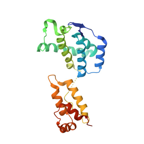
    Retroviruses depend on self-assembly of their capsid proteins (core particle) to yield infectious mature virions. Despite the essential role of the retroviral core, its high polymorphism has hindered high-resolution structural analyses. Here, we report the x-ray structure of the native capsid (CA) protein from bovine leukemia virus. CA is organized as hexamers that deviate substantially from sixfold symmetry, yet adjust to make two-dimensional pseudohexagonal arrays that mimic mature retroviral cores. Intra- and interhexameric quasi-equivalent contacts are uncovered, with flexible trimeric lateral contacts among hexamers, yet preserving very similar dimeric interfaces making the lattice. The conformation of each capsid subunit in the hexamer is therefore dictated by long-range interactions, revealing how the hexamers can also assemble into closed core particles, a relevant feature of retrovirus biology.


  • Organizational Affiliation
    • Institut Pasteur de Montevideo, Unit of Protein Biophysics, Mataojo 2020, 11400, Montevideo, Uruguay. Departamento de Inmunobiología, Facultad de Medicina, Universidad de la República, Avenida General Flores 2125, 11800, Montevideo, Uruguay.

Macromolecule Content 

  • Total Structure Weight: 141.62 kDa 
  • Atom Count: 9,078 
  • Modeled Residue Count: 1,178 
  • Deposited Residue Count: 1,290 
  • Unique protein chains: 1

Macromolecules

Find similar proteins by:|  3D Structure
Entity ID: 1
MoleculeChains  Sequence LengthOrganismDetailsImage
BLV capsid
A, B, C, D, E
A, B, C, D, E, F
215Bovine leukemia virusMutation(s): 0 
Gene Names: gag-pro-pol
UniProt
Find proteins for A7KWZ1 (Bovine leukemia virus)
Explore A7KWZ1 
Go to UniProtKB:  A7KWZ1
Entity Groups
Sequence Clusters30% Identity50% Identity70% Identity90% Identity95% Identity100% Identity
UniProt GroupA7KWZ1
Sequence Annotations
Expand
Reference Sequence

Experimental Data & Validation

Experimental Data

  • Method: X-RAY DIFFRACTION
  • Resolution: 2.75 Å
  • R-Value Free:  0.221 (Depositor), 0.229 (DCC) 
  • R-Value Work:  0.180 (Depositor), 0.182 (DCC) 
  • R-Value Observed: 0.187 (Depositor) 
Space Group: P 65
Unit Cell:
Length ( Å )Angle ( ˚ )
a = 94.28α = 90
b = 94.28β = 90
c = 257.25γ = 120
Software Package:
Software NamePurpose
XDSdata scaling
PDB_EXTRACTdata extraction
PHENIXrefinement

Structure Validation

View Full Validation Report


Entry History 

& Funding Information

Deposition Data


Funding OrganizationLocationGrant Number
Centre National de la Recherche Scientifique (CNRS)FranceLaboratoire International Associe LIA 316
Mercosur Structural Biology Center (CeBEM)Uruguay--
Mercosur/FOCEMUruguayCOF 03/11

Revision History  (Full details and data files)

  • Version 1.0: 2015-06-10
    Type: Initial release
  • Version 1.1: 2015-06-17
    Changes: Database references
  • Version 1.2: 2015-07-15
    Changes: Database references
  • Version 1.3: 2017-11-22
    Changes: Database references, Derived calculations, Other, Refinement description, Source and taxonomy, Structure summary
  • Version 1.4: 2020-01-08
    Changes: Author supporting evidence
  • Version 1.5: 2023-12-27
    Changes: Data collection, Database references, Refinement description