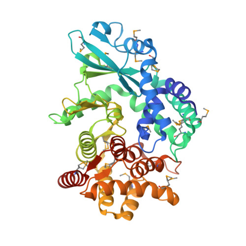
4PED

Mitochondrial ADCK3 employs an atypical protein kinase-like fold to enable coenzyme Q biosynthes


Experimental Data Snapshot

  • Method: X-RAY DIFFRACTION
  • Resolution: 1.64 Å
  • R-Value Free: 0.211 
  • R-Value Work: 0.165 
  • R-Value Observed: 0.167 

wwPDB Validation   3D Report Full Report


This is version 1.7 of the entry. See complete history


Literature

Mitochondrial ADCK3 Employs an Atypical Protein Kinase-like Fold to Enable Coenzyme Q Biosynthesis.

Stefely, J.A.Reidenbach, A.G.Ulbrich, A.Oruganty, K.Floyd, B.J.Jochem, A.Saunders, J.M.Johnson, I.E.Minogue, C.E.Wrobel, R.L.Barber, G.E.Lee, D.Li, S.Kannan, N.Coon, J.J.Bingman, C.A.Pagliarini, D.J.

(2015) Mol Cell 57: 83-94

  • DOI: https://doi.org/10.1016/j.molcel.2014.11.002
  • Primary Citation of Related Structures:  
    4PED

  • PubMed Abstract: 

    The ancient UbiB protein kinase-like family is involved in isoprenoid lipid biosynthesis and is implicated in human diseases, but demonstration of UbiB kinase activity has remained elusive for unknown reasons. Here, we quantitatively define UbiB-specific sequence motifs and reveal their positions within the crystal structure of a UbiB protein, ADCK3. We find that multiple UbiB-specific features are poised to inhibit protein kinase activity, including an N-terminal domain that occupies the typical substrate binding pocket and a unique A-rich loop that limits ATP binding by establishing an unusual selectivity for ADP. A single alanine-to-glycine mutation of this loop flips this coenzyme selectivity and enables autophosphorylation but inhibits coenzyme Q biosynthesis in vivo, demonstrating functional relevance for this unique feature. Our work provides mechanistic insight into UbiB enzyme activity and establishes a molecular foundation for further investigation of how UbiB family proteins affect diseases and diverse biological pathways.


  • Organizational Affiliation

    Department of Biochemistry, University of Wisconsin-Madison, Madison, WI 53706, USA.


Macromolecules
Find similar proteins by:  (by identity cutoff)  |  3D Structure
Entity ID: 1
MoleculeChains Sequence LengthOrganismDetailsImage
Chaperone activity of bc1 complex-like, mitochondrial393Homo sapiensMutation(s): 0 
Gene Names: ADCK3CABC1PP265
EC: 2.7.11 (PDB Primary Data), 2.7 (UniProt)
UniProt & NIH Common Fund Data Resources
Find proteins for Q8NI60 (Homo sapiens)
Explore Q8NI60 
Go to UniProtKB:  Q8NI60
PHAROS:  Q8NI60
GTEx:  ENSG00000163050 
Entity Groups  
Sequence Clusters30% Identity50% Identity70% Identity90% Identity95% Identity100% Identity
UniProt GroupQ8NI60
Sequence Annotations
Expand
  • Reference Sequence
Small Molecules
Modified Residues  1 Unique
IDChains TypeFormula2D DiagramParent
MSE
Query on MSE
A
L-PEPTIDE LINKINGC5 H11 N O2 SeMET
Experimental Data & Validation

Experimental Data

  • Method: X-RAY DIFFRACTION
  • Resolution: 1.64 Å
  • R-Value Free: 0.211 
  • R-Value Work: 0.165 
  • R-Value Observed: 0.167 
  • Space Group: C 1 2 1
Unit Cell:
Length ( Å )Angle ( ˚ )
a = 148.671α = 90
b = 54.557β = 94
c = 45.009γ = 90
Software Package:
Software NamePurpose
HKL-2000data scaling
PHENIXphasing
ARPmodel building
PHENIXmodel building
PHENIXrefinement

Structure Validation

View Full Validation Report


Entry History & Funding Information

Deposition Data


Funding OrganizationLocationGrant Number
National Institutes of Health/National Institute of General Medical Sciences (NIH/NIGMS)United StatesU01GM094622

Revision History  (Full details and data files)

  • Version 1.0: 2014-11-19
    Type: Initial release
  • Version 1.1: 2015-01-14
    Changes: Database references
  • Version 1.2: 2015-01-21
    Changes: Database references
  • Version 1.3: 2015-02-04
    Changes: Derived calculations
  • Version 1.4: 2017-09-06
    Changes: Author supporting evidence, Database references, Derived calculations, Other, Source and taxonomy
  • Version 1.5: 2019-12-25
    Changes: Author supporting evidence
  • Version 1.6: 2023-12-27
    Changes: Data collection, Database references
  • Version 1.7: 2024-11-13
    Changes: Structure summary