4PD1

Structure of gephyrin E domain with Glycine-beta receptor peptide


Experimental Data Snapshot

  • Method: X-RAY DIFFRACTION
  • Resolution: 1.98 Å
  • R-Value Free: 0.196 
  • R-Value Work: 0.159 
  • R-Value Observed: 0.161 

wwPDB Validation   3D Report Full Report


This is version 1.6 of the entry. See complete history


Literature

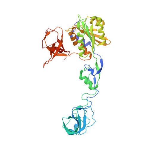
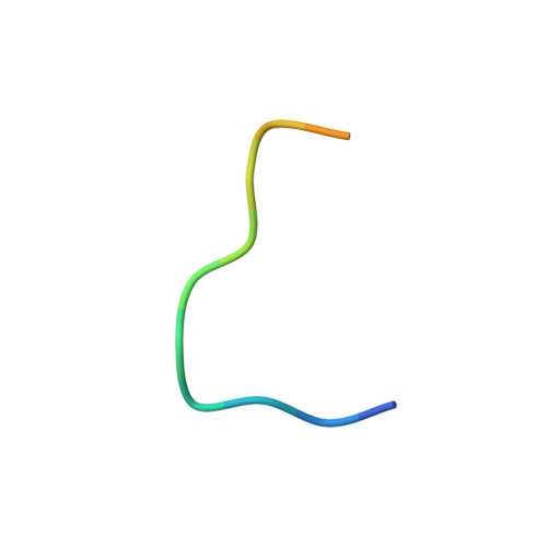
Modulation of gephyrin-glycine receptor affinity by multivalency.

Maric, H.M.Kasaragod, V.B.Schindelin, H.

(2014) ACS Chem Biol 9: 2554-2562

  • DOI: https://doi.org/10.1021/cb500303a
  • Primary Citation of Related Structures:  
    4PD0, 4PD1

  • PubMed Abstract: 

    Gephyrin is a major determinant for the accumulation and anchoring of glycine receptors (GlyRs) and the majority of γ-aminobutyric acid type A receptors (GABAARs) at postsynaptic sites. Here we explored the interaction of gephyrin with a dimeric form of a GlyR β-subunit receptor-derived peptide. A 2 Å crystal structure of the C-terminal domain of gephyrin (GephE) in complex with a 15-residue peptide derived from the GlyR β-subunit defined the core binding site, which we targeted with the dimeric peptide. Biophysical analyses via differential scanning calorimetry (DSC), thermofluor, and isothermal titration calorimetry (ITC) demonstrated that this dimeric ligand is capable of binding simultaneously to two receptor binding sites and that this multivalency results in a 25-fold enhanced affinity. Our study therefore suggests that the oligomeric state of gephyrin and the number of gephyrin-binding subunits in the pentameric GABAARs and GlyRs together control postsynaptic receptor clustering.


  • Organizational Affiliation

    Institute of Structural Biology, Rudolf Virchow Center for Experimental Biomedicine, University of Würzburg , Josef-Schneider-Str. 2, 97080 Würzburg, Germany.


Macromolecules
Find similar proteins by:  (by identity cutoff)  |  3D Structure
Entity ID: 1
MoleculeChains Sequence LengthOrganismDetailsImage
Gephyrin419Rattus norvegicusMutation(s): 0 
Gene Names: GphnGph
EC: 2.7.7.75 (PDB Primary Data), 2.10.1.1 (PDB Primary Data)
UniProt
Find proteins for Q03555 (Rattus norvegicus)
Explore Q03555 
Go to UniProtKB:  Q03555
Entity Groups  
Sequence Clusters30% Identity50% Identity70% Identity90% Identity95% Identity100% Identity
UniProt GroupQ03555
Sequence Annotations
Expand
  • Reference Sequence

Find similar proteins by:  Sequence   |   3D Structure  

Entity ID: 2
MoleculeChains Sequence LengthOrganismDetailsImage
Glycine receptor subunit betaB [auth C]15Rattus norvegicusMutation(s): 0 
UniProt
Find proteins for P20781 (Rattus norvegicus)
Explore P20781 
Go to UniProtKB:  P20781
Entity Groups  
Sequence Clusters30% Identity50% Identity70% Identity90% Identity95% Identity100% Identity
UniProt GroupP20781
Sequence Annotations
Expand
  • Reference Sequence
Experimental Data & Validation

Experimental Data

  • Method: X-RAY DIFFRACTION
  • Resolution: 1.98 Å
  • R-Value Free: 0.196 
  • R-Value Work: 0.159 
  • R-Value Observed: 0.161 
  • Space Group: I 2 2 2
Unit Cell:
Length ( Å )Angle ( ˚ )
a = 85.66α = 90
b = 100.06β = 90
c = 117.5γ = 90
Software Package:
Software NamePurpose
PHENIXrefinement

Structure Validation

View Full Validation Report


Entry History & Funding Information

Deposition Data


Funding OrganizationLocationGrant Number
German Research Foundation (DFG)GermanySchi 425/ 8-1

Revision History  (Full details and data files)

  • Version 1.0: 2014-08-27
    Type: Initial release
  • Version 1.1: 2014-09-03
    Changes: Database references
  • Version 1.2: 2014-09-17
    Changes: Database references
  • Version 1.3: 2014-12-03
    Changes: Database references
  • Version 1.4: 2017-09-27
    Changes: Advisory, Author supporting evidence, Derived calculations, Other, Source and taxonomy, Structure summary
  • Version 1.5: 2020-01-08
    Changes: Author supporting evidence
  • Version 1.6: 2023-09-27
    Changes: Data collection, Database references, Refinement description