4PCY

CRYSTAL STRUCTURE ANALYSES OF REDUCED (CUI) POPLAR PLASTOCYANIN AT SIX PH VALUES


Experimental Data Snapshot

  • Method: X-RAY DIFFRACTION
  • Resolution: 2.15 Å
  • R-Value Observed: 0.150 

wwPDB Validation   3D Report Full Report


This is version 1.3 of the entry. See complete history


Literature

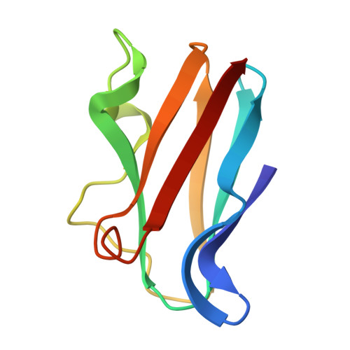
Crystal structure analyses of reduced (CuI) poplar plastocyanin at six pH values.

Guss, J.M.Harrowell, P.R.Murata, M.Norris, V.A.Freeman, H.C.

(1986) J Mol Biol 192: 361-387

  • DOI: 10.1016/0022-2836(86)90371-2
  • Primary Citation of Related Structures:  
    4PCY, 5PCY, 6PCY

  • PubMed Abstract: 
  • The structure of poplar plastocyanin in the reduced (CuI) state has been determined and refined, using counter data recorded from crystals at pH 3.8, 4.4, 5.1, 5.9, 7.0 and 7.8 (resolution 1.9 A, 1.9 A, 2.05 A, 1.7 A, 1.8 A and 2.15 A; the final residual R value was 0 ...

    The structure of poplar plastocyanin in the reduced (CuI) state has been determined and refined, using counter data recorded from crystals at pH 3.8, 4.4, 5.1, 5.9, 7.0 and 7.8 (resolution 1.9 A, 1.9 A, 2.05 A, 1.7 A, 1.8 A and 2.15 A; the final residual R value was 0.15, 0.15, 0.16, 0.17, 0.16 and 0.15, respectively). The molecular and crystal structure of the protein is substantially the same in the reduced state as in the oxidized state. The refinements of the structures of the six forms of the reduced protein could therefore be commenced with a model derived from the known structure of CuII-plastocyanin. The refinements were made by reciprocal space least-squares calculations interspersed with inspections of electron-density difference maps. Precautions were taken to minimize any bias of the results of the refinements in the direction of the starting model. The most significant differences among the structures of the reduced protein at the six pH values, or between them and the structure of the oxidized protein, are concentrated at the Cu site. In the reduced protein at high pH (pH 7.8), the CuI atom is co-ordinated by the N delta(imidazole) atoms of His37 and His87, the S gamma(thiolate) atom of Cys84, and the S delta(thioether) atom of Met92, just as in CuII-plastocyanin. The distorted tetrahedral geometry and the unusually long Cu-S(Met92) bond are retained. The only effects of the change in oxidation state are a lengthening of the two Cu-N(His) bonds by about 0.1 A, and small changes in two bond angles involving the Cu-S(Cys) bond. The high-pH form of reduced plastocyanin accordingly meets all the requirements for efficient electron transfer. As the pH is lowered, the Cu atom and the four Cu-binding protein side-chains appear to undergo small but concerted movements in relation to the rest of the molecule. At low pH (pH 3.8), the CuI atom is trigonally co-ordinated by N delta(His37), S gamma(Cys84) and S delta(Met92). The fourth Cu-ligand bond is broken, the Cu atom making only a van der Waals' contact with the imidazole ring of His87. The trigonal geometry of the Cu atom strongly favours CuI, so that this form of the protein should be redox-inactive. This is known to be the case.(ABSTRACT TRUNCATED AT 400 WORDS)


    Related Citations: 
    • The Crystal Structure of Mercury-Substituted Poplar Plastocyanin at 1.9-Angstroms Resolution
      Church, W.B., Guss, J.M., Potter, J.J., Freeman, H.C.
      (1986) J Biol Chem 261: 234
    • The Crystal Structure of Poplar Apoplastocyanin at 1.8-Angstroms Resolution. The Geometry of the Copper-Binding Site is Created by the Polypeptide
      Garrett, T.P.J., Clingeleffer, D.J., Guss, J.M., Rogers, S.J., Freeman, H.C.
      (1984) J Biol Chem 259: 2822
    • Structure of Oxidized Poplar Plastocyanin at 1.6 Angstroms Resolution
      Guss, J.M., Freeman, H.C.
      (1983) J Mol Biol 169: 521
    • X-Ray Crystal Structure Analysis of Plastocyanin at 2.7 Angstroms Resolution
      Colman, P.M., Freeman, H.C., Guss, J.M., Murata, M., Norris, V.A., Ramshaw, J.A.M., Venkatappa, M.P.
      (1978) Nature 272: 319

    Organizational Affiliation

    Center for Research on Biological Clocks, Texas A&M University College Station, Texas 77843, USA.


Macromolecules
Find similar proteins by:  (by identity cutoff)  |  3D Structure
Entity ID: 1
MoleculeChainsSequence LengthOrganismDetailsImage
PLASTOCYANINA99Populus nigraMutation(s): 0 
Gene Names: PETE
UniProt
Find proteins for P00299 (Populus nigra)
Explore P00299 
Go to UniProtKB:  P00299
Entity Groups  
Sequence Clusters30% Identity50% Identity70% Identity90% Identity95% Identity100% Identity
UniProt GroupP00299
Protein Feature View
Expand
  • Reference Sequence
Small Molecules
Ligands 1 Unique
IDChainsName / Formula / InChI Key2D Diagram3D Interactions
CU
Query on CU

Download Ideal Coordinates CCD File 
B [auth A]COPPER (II) ION
Cu
JPVYNHNXODAKFH-UHFFFAOYSA-N
 Ligand Interaction
Experimental Data & Validation

Experimental Data

  • Method: X-RAY DIFFRACTION
  • Resolution: 2.15 Å
  • R-Value Observed: 0.150 
  • Space Group: P 21 21 21
Unit Cell:
Length ( Å )Angle ( ˚ )
a = 29.72α = 90
b = 47β = 90
c = 57.72γ = 90
Software Package:
Software NamePurpose
PROLSQrefinement

Structure Validation

View Full Validation Report


Entry History 

Deposition Data

Revision History  (Full details and data files)

  • Version 1.0: 1987-01-15
    Type: Initial release
  • Version 1.1: 2008-03-25
    Changes: Version format compliance
  • Version 1.2: 2011-07-13
    Changes: Version format compliance
  • Version 1.3: 2017-11-29
    Changes: Derived calculations, Other