4P99 | pdb_00004p99

Ca2+-stabilized adhesin helps an Antarctic bacterium reach out and bind ice


Experimental Data Snapshot

  • Method: X-RAY DIFFRACTION
  • Resolution: 1.80 Å
  • R-Value Free: 
    0.257 (Depositor), 0.256 (DCC) 
  • R-Value Work: 
    0.221 (Depositor), 0.221 (DCC) 
  • R-Value Observed: 
    0.222 (Depositor) 

wwPDB Validation 3D Report Full Report

Validation slider image for 4P99

Ligand Structure Quality Assessment 


This is version 1.5 of the entry. See complete history

Literature

Ca2+-stabilized adhesin helps an Antarctic bacterium reach out and bind ice.

Vance, T.D.Olijve, L.L.Campbell, R.L.Voets, I.K.Davies, P.L.Guo, S.

(2014) Biosci Rep 34

  • DOI: https://doi.org/10.1042/BSR20140083
  • Primary Citation Related Structures: 
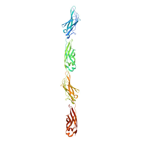
    4P99

  • PubMed Abstract: 

    The large size of a 1.5-MDa ice-binding adhesin [MpAFP (Marinomonas primoryensis antifreeze protein)] from an Antarctic Gram-negative bacterium, M. primoryensis, is mainly due to its highly repetitive RII (Region II). MpAFP_RII contains roughly 120 tandem copies of an identical 104-residue repeat. We have previously determined that a single RII repeat folds as a Ca2+-dependent immunoglobulin-like domain. Here, we solved the crystal structure of RII tetra-tandemer (four tandem RII repeats) to a resolution of 1.8 Å. The RII tetra-tandemer reveals an extended (~190-Å × ~25-Å), rod-like structure with four RII-repeats aligned in series with each other. The inter-repeat regions of the RII tetra-tandemer are strengthened by Ca2+ bound to acidic residues. SAXS (small-angle X-ray scattering) profiles indicate the RII tetra-tandemer is significantly rigidified upon Ca2+ binding, and that the protein's solution structure is in excellent agreement with its crystal structure. We hypothesize that >600 Ca2+ help rigidify the chain of ~120 104-residue repeats to form a ~0.6 μm rod-like structure in order to project the ice-binding domain of MpAFP away from the bacterial cell surface. The proposed extender role of RII can help the strictly aerobic, motile bacterium bind ice in the upper reaches of the Antarctic lake where oxygen and nutrients are most abundant. Ca2+-induced rigidity of tandem Ig-like repeats in large adhesins might be a general mechanism used by bacteria to bind to their substrates and help colonize specific niches.


  • Organizational Affiliation
    • *Protein Function Discovery Group and the Department of Biomedical and Molecular Sciences, Queen's University, Kingston, Ontario, Canada.

Macromolecule Content 

  • Total Structure Weight: 178.38 kDa 
  • Atom Count: 13,555 
  • Modeled Residue Count: 1,660 
  • Deposited Residue Count: 1,748 
  • Unique protein chains: 1

Macromolecules

Find similar proteins by:|  3D Structure
Entity ID: 1
MoleculeChains  Sequence LengthOrganismDetailsImage
MpAFP_RII tetra-tandemer
A, B, C, D
437Marinomonas primoryensisMutation(s): 0 
UniProt
Find proteins for A0A075B5G5 (Marinomonas primoryensis)
Explore A0A075B5G5 
Go to UniProtKB:  A0A075B5G5
Entity Groups
Sequence Clusters30% Identity50% Identity70% Identity90% Identity95% Identity100% Identity
UniProt GroupA0A075B5G5
Sequence Annotations
Expand
Reference Sequence

Small Molecules

Ligands 4 Unique
IDChains Name / Formula / InChI Key2D Diagram3D Interactions
BOG

Query on BOG


Download:Ideal Coordinates CCD File
DC [auth C],
QD [auth C],
ZE [auth D]
octyl beta-D-glucopyranoside
C14 H28 O6
HEGSGKPQLMEBJL-RKQHYHRCSA-N
TRS

Query on TRS


Download:Ideal Coordinates CCD File
PD [auth C]2-AMINO-2-HYDROXYMETHYL-PROPANE-1,3-DIOL
C4 H12 N O3
LENZDBCJOHFCAS-UHFFFAOYSA-O
EDO

Query on EDO


Download:Ideal Coordinates CCD File
CA [auth A]
DA [auth A]
EA [auth A]
FA [auth A]
GA [auth A]
CA [auth A],
DA [auth A],
EA [auth A],
FA [auth A],
GA [auth A],
GD [auth C],
HA [auth A],
HD [auth C],
IA [auth A],
ID [auth C],
JA [auth A],
JD [auth C],
KB [auth B],
KD [auth C],
LA [auth A],
LB [auth B],
LD [auth C],
MA [auth A],
MB [auth B],
MD [auth C],
NB [auth B],
ND [auth C],
OB [auth B],
OD [auth C],
PB [auth B],
QB [auth B],
QE [auth D],
RB [auth B],
RD [auth D],
RE [auth D],
SB [auth B],
SE [auth D],
TB [auth B],
TE [auth D],
UB [auth B],
UE [auth D],
VB [auth B],
VE [auth D],
WB [auth B],
WE [auth D],
XB [auth B],
XE [auth D],
YB [auth B],
YE [auth D]
1,2-ETHANEDIOL
C2 H6 O2
LYCAIKOWRPUZTN-UHFFFAOYSA-N
CA

Query on CA


Download:Ideal Coordinates CCD File
AA [auth A]
AB [auth B]
AC [auth B]
AD [auth C]
AE [auth D]
AA [auth A],
AB [auth B],
AC [auth B],
AD [auth C],
AE [auth D],
BA [auth A],
BB [auth B],
BC [auth C],
BD [auth C],
BE [auth D],
CB [auth B],
CC [auth C],
CD [auth C],
CE [auth D],
DB [auth B],
DD [auth C],
DE [auth D],
E [auth A],
EB [auth B],
EC [auth C],
ED [auth C],
EE [auth D],
F [auth A],
FB [auth B],
FC [auth C],
FD [auth C],
FE [auth D],
G [auth A],
GB [auth B],
GC [auth C],
GE [auth D],
H [auth A],
HB [auth B],
HC [auth C],
HE [auth D],
I [auth A],
IB [auth B],
IC [auth C],
IE [auth D],
J [auth A],
JB [auth B],
JC [auth C],
JE [auth D],
K [auth A],
KA [auth A],
KC [auth C],
KE [auth D],
L [auth A],
LC [auth C],
LE [auth D],
M [auth A],
MC [auth C],
ME [auth D],
N [auth A],
NA [auth B],
NC [auth C],
NE [auth D],
O [auth A],
OA [auth B],
OC [auth C],
OE [auth D],
P [auth A],
PA [auth B],
PC [auth C],
PE [auth D],
Q [auth A],
QA [auth B],
QC [auth C],
R [auth A],
RA [auth B],
RC [auth C],
S [auth A],
SA [auth B],
SC [auth C],
SD [auth D],
T [auth A],
TA [auth B],
TC [auth C],
TD [auth D],
U [auth A],
UA [auth B],
UC [auth C],
UD [auth D],
V [auth A],
VA [auth B],
VC [auth C],
VD [auth D],
W [auth A],
WA [auth B],
WC [auth C],
WD [auth D],
X [auth A],
XA [auth B],
XC [auth C],
XD [auth D],
Y [auth A],
YA [auth B],
YC [auth C],
YD [auth D],
Z [auth A],
ZA [auth B],
ZB [auth B],
ZC [auth C],
ZD [auth D]
CALCIUM ION
Ca
BHPQYMZQTOCNFJ-UHFFFAOYSA-N

Experimental Data & Validation

Experimental Data

  • Method: X-RAY DIFFRACTION
  • Resolution: 1.80 Å
  • R-Value Free:  0.257 (Depositor), 0.256 (DCC) 
  • R-Value Work:  0.221 (Depositor), 0.221 (DCC) 
  • R-Value Observed: 0.222 (Depositor) 
Space Group: P 1
Unit Cell:
Length ( Å )Angle ( ˚ )
a = 47.464α = 90.04
b = 47.469β = 90.01
c = 191.161γ = 90.02
Software Package:
Software NamePurpose
PHENIXrefinement

Structure Validation

View Full Validation Report


Ligand Structure Quality Assessment 


Entry History 

& Funding Information

Deposition Data


Funding OrganizationLocationGrant Number
Canadian Institutes of Health Research (CIHR)Canada394878

Revision History  (Full details and data files)

  • Version 1.0: 2014-06-18
    Type: Initial release
  • Version 1.1: 2014-10-01
    Changes: Database references
  • Version 1.2: 2015-02-04
    Changes: Derived calculations
  • Version 1.3: 2020-01-08
    Changes: Advisory, Author supporting evidence, Derived calculations, Other, Source and taxonomy, Structure summary
  • Version 1.4: 2020-07-29
    Type: Remediation
    Reason: Carbohydrate remediation
    Changes: Data collection, Derived calculations, Refinement description, Structure summary
  • Version 1.5: 2023-12-27
    Changes: Data collection, Database references, Structure summary