4P7A

Crystal Structure of human MLH1


Experimental Data Snapshot

  • Method: X-RAY DIFFRACTION
  • Resolution: 2.30 Å
  • R-Value Free: 0.254 
  • R-Value Work: 0.203 
  • R-Value Observed: 0.205 

wwPDB Validation   3D Report Full Report


Ligand Structure Quality Assessment 


This is version 2.0 of the entry. See complete history

Re-refinement Note

This entry reflects an alternative modeling of the original data in: 3NA3


Literature

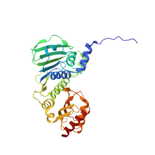
Structure of the human MLH1 N-terminus: implications for predisposition to Lynch syndrome.

Wu, H.Zeng, H.Lam, R.Tempel, W.Kerr, I.D.Min, J.

(2015) Acta Crystallogr F Struct Biol Commun 71: 981-985

  • DOI: https://doi.org/10.1107/S2053230X15010183
  • Primary Citation of Related Structures:  
    4P7A

  • PubMed Abstract: 
  • Mismatch repair prevents the accumulation of erroneous insertions/deletions and non-Watson-Crick base pairs in the genome. Pathogenic mutations in the MLH1 gene are associated with a predisposition to Lynch and Turcot's syndromes. Although genetic testing for these mutations is available, robust classification of variants requires strong clinical and functional support ...

    Mismatch repair prevents the accumulation of erroneous insertions/deletions and non-Watson-Crick base pairs in the genome. Pathogenic mutations in the MLH1 gene are associated with a predisposition to Lynch and Turcot's syndromes. Although genetic testing for these mutations is available, robust classification of variants requires strong clinical and functional support. Here, the first structure of the N-terminus of human MLH1, determined by X-ray crystallography, is described. The structure shares a high degree of similarity with previously determined prokaryotic MLH1 homologs; however, this structure affords a more accurate platform for the classification of MLH1 variants.


    Organizational Affiliation

    Structural Genomics Consortium, University of Toronto, 101 College Street, Toronto, ON M5G 1L7, Canada.


Macromolecules
Find similar proteins by:  (by identity cutoff)  |  3D Structure
Entity ID: 1
MoleculeChainsSequence LengthOrganismDetailsImage
DNA mismatch repair protein Mlh1348Homo sapiensMutation(s): 0 
Gene Names: MLH1COCA2
UniProt & NIH Common Fund Data Resources
Find proteins for P40692 (Homo sapiens)
Explore P40692 
Go to UniProtKB:  P40692
PHAROS:  P40692
Entity Groups  
Sequence Clusters30% Identity50% Identity70% Identity90% Identity95% Identity100% Identity
UniProt GroupP40692
Protein Feature View
Expand
  • Reference Sequence
Experimental Data & Validation

Experimental Data

  • Method: X-RAY DIFFRACTION
  • Resolution: 2.30 Å
  • R-Value Free: 0.254 
  • R-Value Work: 0.203 
  • R-Value Observed: 0.205 
  • Space Group: P 64
Unit Cell:
Length ( Å )Angle ( ˚ )
a = 94.568α = 90
b = 94.568β = 90
c = 85.825γ = 120
Software Package:
Software NamePurpose
REFMACrefinement
Aimlessdata scaling
BUSTERrefinement
XDSdata reduction
PDB_EXTRACTdata extraction

Structure Validation

View Full Validation Report


Ligand Structure Quality Assessment 


Entry History 

Deposition Data

Revision History  (Full details and data files)

  • Version 1.0: 2014-04-09
    Type: Initial release
  • Version 1.1: 2014-11-12
    Changes: Other
  • Version 1.2: 2015-12-16
    Changes: Database references
  • Version 2.0: 2019-07-17
    Changes: Advisory, Atomic model, Data collection, Derived calculations, Refinement description, Structure summary