4P3C | pdb_00004p3c

MT1-MMP:Fab complex (Form I)


Experimental Data Snapshot

  • Method: X-RAY DIFFRACTION
  • Resolution: 1.94 Å
  • R-Value Free: 
    0.200 (Depositor), 0.201 (DCC) 
  • R-Value Work: 
    0.152 (Depositor), 0.154 (DCC) 
  • R-Value Observed: 
    0.156 (Depositor) 

Starting Model: experimental
View more details

wwPDB Validation 3D Report Full Report

Validation slider image for 4P3C

This is version 1.6 of the entry. See complete history

Literature

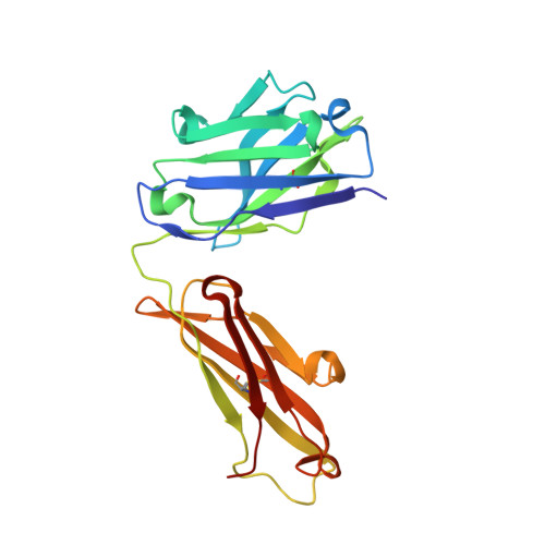
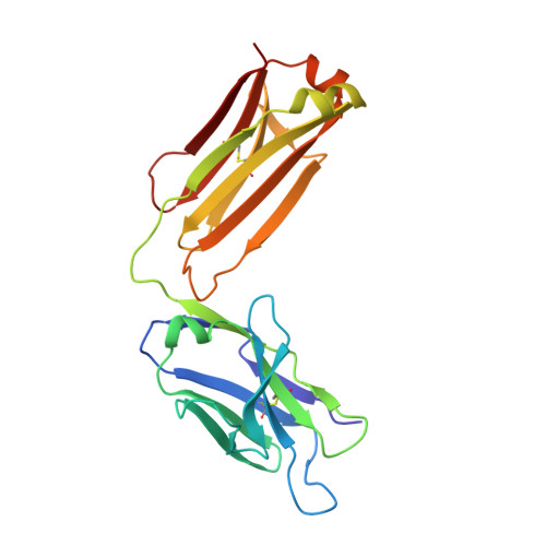
Inhibition mechanism of membrane metalloprotease by an exosite-swiveling conformational antibody.

Udi, Y.Grossman, M.Solomonov, I.Dym, O.Rozenberg, H.Moreno, V.Cuniasse, P.Dive, V.Arroyo, A.G.Sagi, I.

(2015) Structure 23: 104-115

  • DOI: https://doi.org/10.1016/j.str.2014.10.012
  • Primary Citation Related Structures: 
    4OUU, 4P3C, 4P3D, 4QXU

  • PubMed Abstract: 

    Membrane type 1 metalloprotease (MT1-MMP) is a membrane-anchored, zinc-dependent protease. MT1-MMP is an important mediator of cell migration and invasion, and overexpression of this enzyme has been correlated with the malignancy of various tumor types. Therefore, modulators of MT1-MMP activity are proposed to possess therapeutic potential in numerous invasive diseases. Here we report the inhibition mode of MT1-MMP by LEM-2/15 antibody, which targets a surface epitope of MT1-MMP. Specifically, the crystal structures of Fab LEM-2/15 in complex with the MT1-MMP surface antigen suggest that conformational swiveling of the enzyme surface loop is required for effective binding and consequent inhibition of MT1-MMP activity on the cell membrane. This inhibition mechanism appears to be effective in controlling active MT1-MMP in endothelial cells and at the leading edge of migratory cancer cells.


  • Organizational Affiliation
    • Department of Biological Regulation, The Weizmann Institute of Science, Rehovot 76100, Israel.

Macromolecule Content 

  • Total Structure Weight: 49.87 kDa 
  • Atom Count: 3,992 
  • Modeled Residue Count: 449 
  • Deposited Residue Count: 449 
  • Unique protein chains: 3

Macromolecules

Find similar proteins by:|  3D Structure
Entity ID: 1
MoleculeChains  Sequence LengthOrganismDetailsImage
Heavy Chain Fab fragment of antibody LEM-2/15A [auth H]218Mus musculusMutation(s): 0 
Find similar proteins by:|  3D Structure
Entity ID: 2
MoleculeChains  Sequence LengthOrganismDetailsImage
Light Chain Fab fragment of antibody LEM-2/15B [auth L]218Mus musculusMutation(s): 0 
Find similar proteins by:  Sequence   |   3D Structure  
Entity ID: 3
MoleculeChains  Sequence LengthOrganismDetailsImage
Matrix metalloproteinase-14C [auth M]13Homo sapiensMutation(s): 0 
Gene Names: MMP14
EC: 3.4.24.80
UniProt & NIH Common Fund Data Resources
Find proteins for P50281 (Homo sapiens)
Explore P50281 
Go to UniProtKB:  P50281
PHAROS:  P50281
GTEx:  ENSG00000157227 
Entity Groups
Sequence Clusters30% Identity50% Identity70% Identity90% Identity95% Identity100% Identity
UniProt GroupP50281
Sequence Annotations
Expand
Reference Sequence

Small Molecules

Ligands 5 Unique
IDChains Name / Formula / InChI Key2D Diagram3D Interactions
EPE

Query on EPE


Download:Ideal Coordinates CCD File
E [auth H]4-(2-HYDROXYETHYL)-1-PIPERAZINE ETHANESULFONIC ACID
C8 H18 N2 O4 S
JKMHFZQWWAIEOD-UHFFFAOYSA-N
EDO

Query on EDO


Download:Ideal Coordinates CCD File
F [auth H]
G [auth H]
H
I [auth H]
J [auth H]
F [auth H],
G [auth H],
H,
I [auth H],
J [auth H],
N [auth L]
1,2-ETHANEDIOL
C2 H6 O2
LYCAIKOWRPUZTN-UHFFFAOYSA-N
ACT

Query on ACT


Download:Ideal Coordinates CCD File
K [auth H],
L [auth H],
O [auth L]
ACETATE ION
C2 H3 O2
QTBSBXVTEAMEQO-UHFFFAOYSA-M
CL

Query on CL


Download:Ideal Coordinates CCD File
D [auth H]CHLORIDE ION
Cl
VEXZGXHMUGYJMC-UHFFFAOYSA-M
MG

Query on MG


Download:Ideal Coordinates CCD File
M [auth L]MAGNESIUM ION
Mg
JLVVSXFLKOJNIY-UHFFFAOYSA-N

Experimental Data & Validation

Experimental Data

  • Method: X-RAY DIFFRACTION
  • Resolution: 1.94 Å
  • R-Value Free:  0.200 (Depositor), 0.201 (DCC) 
  • R-Value Work:  0.152 (Depositor), 0.154 (DCC) 
  • R-Value Observed: 0.156 (Depositor) 
Space Group: P 2 21 21
Unit Cell:
Length ( Å )Angle ( ˚ )
a = 52.314α = 90
b = 80.975β = 90
c = 95.568γ = 90
Software Package:
Software NamePurpose
DNAdata collection
HKL-2000data scaling
PHASERphasing
PHENIXrefinement

Structure Validation

View Full Validation Report


Entry History 

Deposition Data

Revision History  (Full details and data files)

  • Version 1.0: 2014-12-17
    Type: Initial release
  • Version 1.1: 2015-01-14
    Changes: Database references
  • Version 1.2: 2015-01-21
    Changes: Database references
  • Version 1.3: 2015-02-18
    Changes: Refinement description
  • Version 1.4: 2017-09-27
    Changes: Advisory, Database references, Derived calculations, Other, Refinement description, Source and taxonomy
  • Version 1.5: 2023-09-27
    Changes: Data collection, Database references, Derived calculations, Refinement description
  • Version 1.6: 2024-10-30
    Changes: Structure summary