4P2G | pdb_00004p2g

Crystal structure of DJ-1 in sulfinic acid form (aged crystal)


Experimental Data Snapshot

  • Method: X-RAY DIFFRACTION
  • Resolution: 1.35 Å
  • R-Value Free: 
    0.120 (Depositor), 0.124 (DCC) 
  • R-Value Work: 
    0.098 (Depositor), 0.103 (DCC) 
  • R-Value Observed: 
    0.099 (Depositor) 

Starting Model: experimental
View more details

wwPDB Validation 3D Report Full Report

Validation slider image for 4P2G

This is version 1.4 of the entry. See complete history

Literature

Thermodynamic and Structural Characterization of the Specific Binding of Zn(II) to Human Protein DJ-1.

Tashiro, S.Caaveiro, J.M.Wu, C.X.Hoang, Q.Q.Tsumoto, K.

(2014) Biochemistry 53: 2218-2220

  • DOI: https://doi.org/10.1021/bi500294h
  • Primary Citation Related Structures: 
    4P2G, 4P34, 4P35, 4P36

  • PubMed Abstract: 

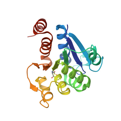
    Mutations of DJ-1 cause familial Parkinson's disease (PD), although the role of DJ-1 in PD remains unresolved. Very recent reports have shown that DJ-1 interacts with copper ions. This evidence opens new avenues to understanding the function of DJ-1 and its role in PD. Herein, we report that Zn(II) binds to DJ-1 with great selectivity among the other metals examined: Mn(II), Fe(II), Co(II), Ni(II), and Cu(II). High-resolution X-ray crystallography (1.18 Å resolution) shows Zn(II) is coordinated to the protein by the key residues Cys106 and Glu18. These results suggest that DJ-1 may be regulated and/or stabilized by Zn(II).


  • Organizational Affiliation
    • Department of Medical Genome Sciences, Graduate School of Frontier Sciences, The University of Tokyo , Kashiwa 277-8562, Japan.

Macromolecule Content 

  • Total Structure Weight: 20.25 kDa 
  • Atom Count: 1,766 
  • Modeled Residue Count: 187 
  • Deposited Residue Count: 189 
  • Unique protein chains: 1

Macromolecules

Find similar proteins by:|  3D Structure
Entity ID: 1
MoleculeChains  Sequence LengthOrganismDetailsImage
Protein DJ-1189Homo sapiensMutation(s): 0 
Gene Names: PARK7
EC: 3.4 (PDB Primary Data), 3.5.1 (UniProt), 3.1.2 (UniProt), 3.5.1.124 (UniProt)
UniProt & NIH Common Fund Data Resources
Find proteins for Q99497 (Homo sapiens)
Explore Q99497 
Go to UniProtKB:  Q99497
PHAROS:  Q99497
GTEx:  ENSG00000116288 
Entity Groups
Sequence Clusters30% Identity50% Identity70% Identity90% Identity95% Identity100% Identity
UniProt GroupQ99497
Sequence Annotations
Expand
Reference Sequence

Small Molecules

Modified Residues  1 Unique
IDChains TypeFormula2D DiagramParent
CSD
Query on CSD
A
L-PEPTIDE LINKINGC3 H7 N O4 SCYS

Experimental Data & Validation

Experimental Data

  • Method: X-RAY DIFFRACTION
  • Resolution: 1.35 Å
  • R-Value Free:  0.120 (Depositor), 0.124 (DCC) 
  • R-Value Work:  0.098 (Depositor), 0.103 (DCC) 
  • R-Value Observed: 0.099 (Depositor) 
Space Group: P 31 2 1
Unit Cell:
Length ( Å )Angle ( ˚ )
a = 74.76α = 90
b = 74.76β = 90
c = 75.03γ = 120
Software Package:
Software NamePurpose
REFMACrefinement

Structure Validation

View Full Validation Report


Entry History 

Deposition Data

Revision History  (Full details and data files)

  • Version 1.0: 2014-04-09
    Type: Initial release
  • Version 1.1: 2014-05-14
    Changes: Database references
  • Version 1.2: 2014-07-23
    Changes: Structure summary
  • Version 1.3: 2023-09-27
    Changes: Advisory, Data collection, Database references, Derived calculations, Other, Refinement description, Source and taxonomy
  • Version 1.4: 2024-11-13
    Changes: Structure summary