Subdomain II of alpha-isopropylmalate synthase is essential for activity: inferring a mechanism of feedback inhibition.
Zhang, Z., Wu, J., Lin, W., Wang, J., Yan, H., Zhao, W., Ma, J., Ding, J., Zhang, P., Zhao, G.P.(2014) J Biol Chem 289: 27966-27978
- PubMed: 25128527 Search on PubMedSearch on PubMed Central
- DOI: https://doi.org/10.1074/jbc.M114.559716
- Primary Citation Related Structures:
4OV4, 4OV9 - PubMed Abstract:
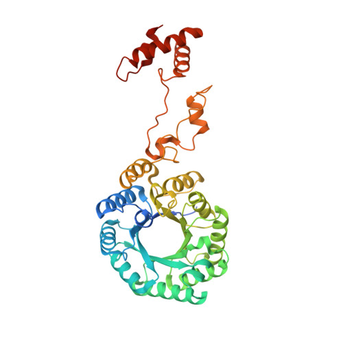
The committed step of leucine biosynthesis, converting acetyl-CoA and α-ketoisovalerate into α-isopropylmalate, is catalyzed by α-isopropylmalate synthase (IPMS), an allosteric enzyme subjected to feedback inhibition by the end product L-leucine. We characterized the short form IPMS from Leptospira biflexa (LbIPMS2), which exhibits a catalytic activity comparable with that of the long form IPMS (LbIPMS1) and has a similar N-terminal domain followed by subdomain I and subdomain II but lacks the whole C-terminal regulatory domain. We found that partial deletion of the regulatory domain of LbIPMS1 resulted in a loss of about 50% of the catalytic activity; however, when the regulatory domain was deleted up to Arg-385, producing a protein that is almost equivalent to the intact LbIPMS2, about 90% of the activity was maintained. Moreover, in LbIPMS2 or LbIPMS1, further deletion of several residues from the C terminus of subdomain II significantly impaired or completely abolished the catalytic activity, respectively. These results define a complete and independently functional catalytic module of IPMS consisting of both the N-terminal domain and the two subdomains. Structural comparison of LbIPMS2 and the Mycobacterium tuberculosis IPMS revealed two different conformations of subdomain II that likely represent two substrate-binding states related to cooperative catalysis. The biochemical and structural analyses together with the previously published hydrogen-deuterium exchange data led us to propose a conformation transition mechanism for feedback inhibition mediated by subdomains I and II that might associated with alteration of the binding affinity toward acetyl-CoA.
- From the Chinese Academy of Sciences Key Laboratory of Synthetic Biology and Shanghai International Travel Healthcare Center, Shanghai Entry-Exit Inspection and Quarantine Bureau, Shanghai 200335, China.
Organizational Affiliation: