4OUJ

Crystal structure of HA33B-Lac


Experimental Data Snapshot

  • Method: X-RAY DIFFRACTION
  • Resolution: 1.46 Å
  • R-Value Free: 0.181 
  • R-Value Work: 0.163 
  • R-Value Observed: 0.164 

wwPDB Validation   3D Report Full Report


This is version 2.1 of the entry. See complete history


Literature

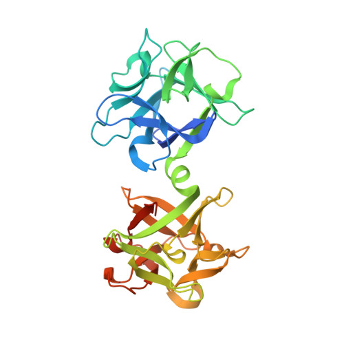
High-resolution crystal structure of HA33 of botulinum neurotoxin type B progenitor toxin complex.

Lee, K.Lam, K.H.Kruel, A.M.Perry, K.Rummel, A.Jin, R.

(2014) Biochem Biophys Res Commun 446: 568-573

  • DOI: https://doi.org/10.1016/j.bbrc.2014.03.008
  • Primary Citation of Related Structures:  
    4OUJ

  • PubMed Abstract: 

    Botulinum neurotoxins (BoNTs) are produced as progenitor toxin complexes (PTCs) by Clostridium botulinum. The PTCs are composed of BoNT and non-toxic neurotoxin-associated proteins (NAPs), which serve to protect and deliver BoNT through the gastrointestinal tract in food borne botulism. HA33 is a key NAP component that specifically recognizes host carbohydrates and helps enrich PTC on the intestinal lumen preceding its transport across the epithelial barriers. Here, we report the crystal structure of HA33 of type B PTC (HA33/B) in complex with lactose at 1.46Å resolution. The structural comparisons among HA33 of serotypes A-D reveal two different HA33-glycan interaction modes. The glycan-binding pockets on HA33/A and B are more suitable to recognize galactose-containing glycans in comparison to the equivalent sites on HA33/C and D. On the contrary, HA33/C and D could potentially recognize Neu5Ac as an independent receptor, whereas HA33/A and B do not. These findings indicate that the different oral toxicity and host susceptibility observed among different BoNT serotypes could be partly determined by the serotype-specific interaction between HA33 and host carbohydrate receptors. Furthermore, we have identified a key structural water molecule that mediates the HA33/B-lactose interactions. It provides the structural basis for development of new receptor-mimicking compounds, which have enhanced binding affinity with HA33 through their water-displacing moiety.


  • Organizational Affiliation

    Department of Physiology and Biophysics, University of California, Irvine, CA 92697, USA.


Macromolecules
Find similar proteins by:  (by identity cutoff)  |  3D Structure
Entity ID: 1
MoleculeChains Sequence LengthOrganismDetailsImage
Hemagglutinin component HA33
A, B
307Clostridium botulinum B1 str. OkraMutation(s): 5 
Gene Names: CLD_A0071ha33
UniProt
Find proteins for B1INP8 (Clostridium botulinum (strain Okra / Type B1))
Explore B1INP8 
Go to UniProtKB:  B1INP8
Entity Groups  
Sequence Clusters30% Identity50% Identity70% Identity90% Identity95% Identity100% Identity
UniProt GroupB1INP8
Sequence Annotations
Expand
  • Reference Sequence
Oligosaccharides

Help

Entity ID: 2
MoleculeChains Length2D Diagram Glycosylation3D Interactions
beta-D-galactopyranose-(1-4)-alpha-D-glucopyranose
C, D
2N/A
Glycosylation Resources
GlyTouCan:  G88362QR
GlyCosmos:  G88362QR
GlyGen:  G88362QR
Biologically Interesting Molecules (External Reference) 1 Unique
Experimental Data & Validation

Experimental Data

  • Method: X-RAY DIFFRACTION
  • Resolution: 1.46 Å
  • R-Value Free: 0.181 
  • R-Value Work: 0.163 
  • R-Value Observed: 0.164 
  • Space Group: P 1 21 1
Unit Cell:
Length ( Å )Angle ( ˚ )
a = 83.819α = 90
b = 36.959β = 102.11
c = 114.624γ = 90
Software Package:
Software NamePurpose
ADSCdata collection
PHASESphasing
PHENIXrefinement
HKL-2000data reduction
HKL-2000data scaling

Structure Validation

View Full Validation Report


Entry History 

Deposition Data

Revision History  (Full details and data files)

  • Version 1.0: 2014-06-04
    Type: Initial release
  • Version 2.0: 2020-07-29
    Type: Remediation
    Reason: Carbohydrate remediation
    Changes: Atomic model, Data collection, Database references, Derived calculations, Non-polymer description, Structure summary
  • Version 2.1: 2023-09-20
    Changes: Data collection, Database references, Refinement description, Structure summary