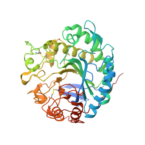
4OOZ

Crystal structure of beta-1,4-D-mannanase from Cryptopygus antarcticus in complex with mannopentaose


Experimental Data Snapshot

  • Method: X-RAY DIFFRACTION
  • Resolution: 2.60 Å
  • R-Value Free: 0.271 
  • R-Value Work: 0.235 
  • R-Value Observed: 0.237 

wwPDB Validation   3D Report Full Report


Ligand Structure Quality Assessment 


This is version 2.0 of the entry. See complete history


Literature

Structure-based investigation into the functional roles of the extended loop and substrate-recognition sites in an endo-beta-1,4-d-mannanase from the Antarctic springtail, Cryptopygus antarcticus.

Kim, M.K.An, Y.J.Song, J.M.Jeong, C.S.Kang, M.H.Kwon, K.K.Lee, Y.H.Cha, S.S.

(2014) Proteins 82: 3217-3223

  • DOI: https://doi.org/10.1002/prot.24655
  • Primary Citation of Related Structures:  
    4OOU, 4OOZ

  • PubMed Abstract: 

    Endo-β-1,4-D-mannanase from the Antarctic springtail, Cryptopygus antarcticus (CaMan), is a cold-adapted β-mannanase that has the lowest optimum temperature (30°C) of all known β-mannanases. Here, we report the apo- and mannopentaose (M5) complex structures of CaMan. Structural comparison of CaMan with other β-mannanases from the multicellular animals reveals that CaMan has an extended loop that alters topography of the active site. Structural and mutational analyses suggest that this extended loop is linked to the cold-adapted enzymatic activity. From the CaMan-M5 complex structure, we defined the mannose-recognition subsites and observed unreported M5 binding site on the surface of CaMan.


  • Organizational Affiliation

    Marine Biotechnology Research Division, Korea Institute of Ocean Science and Technology, Ansan, 426-744, Republic of Korea.


Macromolecules
Find similar proteins by:  (by identity cutoff)  |  3D Structure
Entity ID: 1
MoleculeChains Sequence LengthOrganismDetailsImage
Beta-1,4-mannanase
A, B
388Cryptopygus antarcticusMutation(s): 0 
EC: 3.2.1.78
UniProt
Find proteins for B4XC07 (Cryptopygus antarcticus)
Explore B4XC07 
Go to UniProtKB:  B4XC07
Entity Groups  
Sequence Clusters30% Identity50% Identity70% Identity90% Identity95% Identity100% Identity
UniProt GroupB4XC07
Sequence Annotations
Expand
  • Reference Sequence
Oligosaccharides

Help

Entity ID: 2
MoleculeChains Length2D Diagram Glycosylation3D Interactions
beta-D-mannopyranose-(1-4)-beta-D-mannopyranose-(1-4)-beta-D-mannopyranose-(1-4)-beta-D-mannopyranose
C, E
4N/A
Glycosylation Resources
GlyTouCan:  G51338PC
GlyCosmos:  G51338PC
GlyGen:  G51338PC
Entity ID: 3
MoleculeChains Length2D Diagram Glycosylation3D Interactions
beta-D-mannopyranose-(1-4)-beta-D-mannopyranose-(1-4)-beta-D-mannopyranose-(1-4)-beta-D-mannopyranose-(1-4)-beta-D-mannopyranose
D, F
5N/A
Glycosylation Resources
GlyTouCan:  G91924WY
GlyCosmos:  G91924WY
GlyGen:  G91924WY
Small Molecules
Experimental Data & Validation

Experimental Data

  • Method: X-RAY DIFFRACTION
  • Resolution: 2.60 Å
  • R-Value Free: 0.271 
  • R-Value Work: 0.235 
  • R-Value Observed: 0.237 
  • Space Group: P 21 21 21
Unit Cell:
Length ( Å )Angle ( ˚ )
a = 74.023α = 90
b = 82.453β = 90
c = 164.324γ = 90
Software Package:
Software NamePurpose
HKL-2000data collection
MOLREPphasing
PHENIXrefinement
HKL-2000data reduction
HKL-2000data scaling

Structure Validation

View Full Validation Report


Ligand Structure Quality Assessment 


Entry History 

Deposition Data

Revision History  (Full details and data files)

  • Version 1.0: 2014-08-06
    Type: Initial release
  • Version 1.1: 2014-11-05
    Changes: Database references
  • Version 2.0: 2020-07-29
    Type: Remediation
    Reason: Carbohydrate remediation
    Changes: Advisory, Atomic model, Data collection, Database references, Derived calculations, Structure summary