


Crystal structure of YagE, a KDG aldolase protein in complex with 2-Keto-3-deoxy gluconate
Manoj Kumar, P., Bhaskar, V., Manicka, S., Krishnaswamy, S.To be published.
Experimental Data Snapshot
Entity ID: 1 | |||||
|---|---|---|---|---|---|
| Molecule | Chains | Sequence Length | Organism | Details | Image |
| Probable 2-keto-3-deoxy-galactonate aldolase YagE | 343 | Escherichia coli K-12 | Mutation(s): 0 Gene Names: b0268, JW0261, yagE EC: 4.1.2 (PDB Primary Data), 4.1.2.51 (UniProt), 4.1.2.28 (UniProt) | 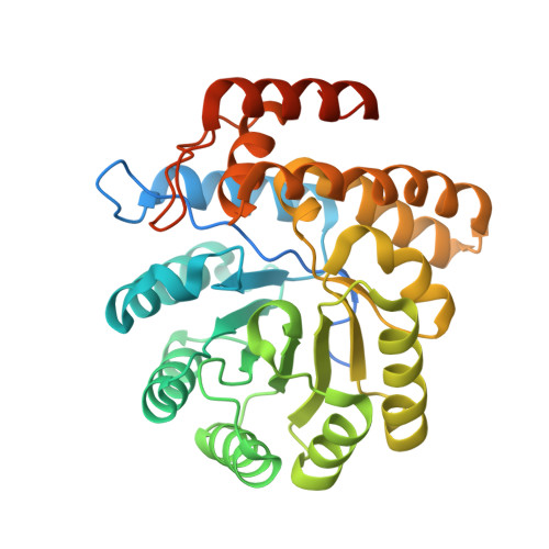 | |
UniProt | |||||
Entity Groups | |||||
| Sequence Clusters | 30% Identity50% Identity70% Identity90% Identity95% Identity100% Identity | ||||
| UniProt Group | P75682 | ||||
Sequence AnnotationsExpand | |||||
Reference Sequence | |||||
| Ligands 3 Unique | |||||
|---|---|---|---|---|---|
| ID | Chains | Name / Formula / InChI Key | 2D Diagram | 3D Interactions | |
| KDG Download:Ideal Coordinates CCD File | AA [auth C], J [auth A], JA [auth D], S [auth B] | 2-KETO-3-DEOXYGLUCONATE C6 H10 O6 WPAMZTWLKIDIOP-WVZVXSGGSA-N |  | ||
| GOL Download:Ideal Coordinates CCD File | EA [auth D] F [auth A] G [auth A] GA [auth D] H [auth A] | GLYCEROL C3 H8 O3 PEDCQBHIVMGVHV-UHFFFAOYSA-N |  | ||
| EDO Download:Ideal Coordinates CCD File | BA [auth D] CA [auth D] DA [auth D] E [auth A] FA [auth D] | 1,2-ETHANEDIOL C2 H6 O2 LYCAIKOWRPUZTN-UHFFFAOYSA-N |  | ||
| Length ( Å ) | Angle ( ˚ ) |
|---|---|
| a = 141.17 | α = 90 |
| b = 155.41 | β = 90 |
| c = 55.51 | γ = 90 |
| Software Name | Purpose |
|---|---|
| XSCALE | data scaling |
| PHASER | phasing |
| REFMAC | refinement |
| PDB_EXTRACT | data extraction |


