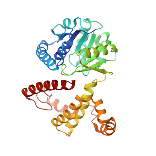
4OM8

Crystal structure of 5-formly-3-hydroxy-2-methylpyridine 4-carboxylic acid (FHMPC) 5-dehydrogenase, an NAD+ dependent dismutase.


Experimental Data Snapshot

  • Method: X-RAY DIFFRACTION
  • Resolution: 1.55 Å
  • R-Value Free: 0.188 
  • R-Value Work: 0.155 
  • R-Value Observed: 0.157 

Starting Model: experimental
View more details

wwPDB Validation   3D Report Full Report


Ligand Structure Quality Assessment 


This is version 1.3 of the entry. See complete history


Literature

Crystal structure of 5-formyl-3-hydroxy-2-methylpyridine 4-carboxylic acid 5-dehydrogenase, an NAD(+)-dependent dismutase from Mesorhizobium loti

Mugo, A.N.Kobayashi, J.Mikami, B.Yoshikane, Y.Yagi, T.Ohnishi, K.

(2015) Biochem Biophys Res Commun 456: 35-40

  • DOI: https://doi.org/10.1016/j.bbrc.2014.11.028
  • Primary Citation of Related Structures:  
    4OM8

  • PubMed Abstract: 

    5-Formyl-3-hydroxy-2-methylpyridine 4-carboxylic acid 5-dehydrogenase (FHMPCDH) from Mesorhizobium loti is the fifth enzyme in degradation pathway I for pyridoxine. The enzyme catalyzes a dismutation reaction: the oxidation of 5-formyl-3-hydroxy-2-methylpyridine 4-carboxylic acid (FHMPC) to 3-hydroxy-2-methylpyridine 4,5-dicarboxylic acid with NAD(+) and reduction of FHMPC to 4-pyridoxic acid with NADH. FHMPCDH belongs to the l-3-hydroxyacyl-CoA dehydrogenase (HAD) family. The crystal structure was determined by molecular replacement and refined to a resolution of 1.55Å (R-factor of 16.4%, Rfree=19.4%). There were two monomers in the asymmetric unit. The overall structure of the monomer consisted of N- and C-terminal domains connected by a short linker loop. The monomer was similar to members of the HAD family (RMSD=1.9Å). The active site was located between the domains and highly conserved to that of human heart l-3-hydroxyacyl-CoA dehydrogenase (HhHAD). His-Glu catalytic dyad, a serine and two asparagine residues of HhHAD were conserved. Ser116, His137 and Glu149 in FHMPCDH are connected by a hydrogen bonding network forming a catalytic triad. The functions of the active site residues in the reaction mechanism are discussed.


  • Organizational Affiliation

    Research Institute of Molecular Genetics, Kochi University, Nankoku, Kochi 783-8502, Japan.


Macromolecules
Find similar proteins by:  (by identity cutoff)  |  3D Structure
Entity ID: 1
MoleculeChains Sequence LengthOrganismDetailsImage
3-hydroxybutyryl-coA dehydrogenase
A, B
309Mesorhizobium japonicum MAFF 303099Mutation(s): 0 
Gene Names: Mesorhizobium loti: mlr6793mlr6793
UniProt
Find proteins for Q988C8 (Mesorhizobium japonicum (strain LMG 29417 / CECT 9101 / MAFF 303099))
Explore Q988C8 
Go to UniProtKB:  Q988C8
Entity Groups  
Sequence Clusters30% Identity50% Identity70% Identity90% Identity95% Identity100% Identity
UniProt GroupQ988C8
Sequence Annotations
Expand
  • Reference Sequence
Small Molecules
Ligands 4 Unique
IDChains Name / Formula / InChI Key2D Diagram3D Interactions
NAD
Query on NAD

Download Ideal Coordinates CCD File 
D [auth A],
H [auth B]
NICOTINAMIDE-ADENINE-DINUCLEOTIDE
C21 H27 N7 O14 P2
BAWFJGJZGIEFAR-NNYOXOHSSA-N
GOL
Query on GOL

Download Ideal Coordinates CCD File 
J [auth B]GLYCEROL
C3 H8 O3
PEDCQBHIVMGVHV-UHFFFAOYSA-N
BME
Query on BME

Download Ideal Coordinates CCD File 
C [auth A],
G [auth B]
BETA-MERCAPTOETHANOL
C2 H6 O S
DGVVWUTYPXICAM-UHFFFAOYSA-N
ACT
Query on ACT

Download Ideal Coordinates CCD File 
E [auth A],
F [auth A],
I [auth B]
ACETATE ION
C2 H3 O2
QTBSBXVTEAMEQO-UHFFFAOYSA-M
Experimental Data & Validation

Experimental Data

  • Method: X-RAY DIFFRACTION
  • Resolution: 1.55 Å
  • R-Value Free: 0.188 
  • R-Value Work: 0.155 
  • R-Value Observed: 0.157 
  • Space Group: C 1 2 1
Unit Cell:
Length ( Å )Angle ( ˚ )
a = 122.805α = 90
b = 44.212β = 116.35
c = 109.343γ = 90
Software Package:
Software NamePurpose
REFMACrefinement
PDB_EXTRACTdata extraction
HKL-2000data collection
HKL-2000data reduction
HKL-2000data scaling
MOLREPphasing

Structure Validation

View Full Validation Report


Ligand Structure Quality Assessment 


Entry History 

Deposition Data

Revision History  (Full details and data files)

  • Version 1.0: 2015-01-28
    Type: Initial release
  • Version 1.1: 2015-02-04
    Changes: Database references
  • Version 1.2: 2015-02-18
    Changes: Other
  • Version 1.3: 2023-11-08
    Changes: Data collection, Database references, Derived calculations, Refinement description