4OLN | pdb_00004oln

Ancestral Steroid Receptor 1 in complex with estrogen response element DNA


Experimental Data Snapshot

  • Method: X-RAY DIFFRACTION
  • Resolution: 1.70 Å
  • R-Value Free: 
    0.206 (Depositor), 0.205 (DCC) 
  • R-Value Work: 
    0.175 (Depositor), 0.176 (DCC) 
  • R-Value Observed: 
    0.176 (Depositor) 

wwPDB Validation 3D Report Full Report

Validation slider image for 4OLN

This is version 1.2 of the entry. See complete history

Literature

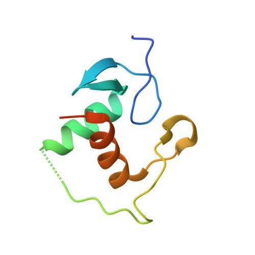
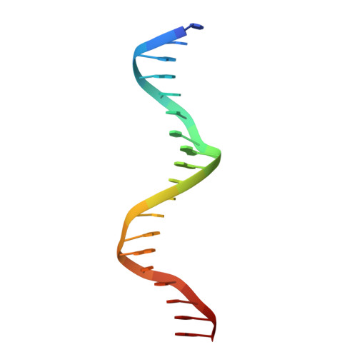
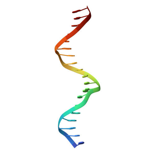
Evolution of DNA specificity in a transcription factor family produced a new gene regulatory module.

McKeown, A.N.Bridgham, J.T.Anderson, D.W.Murphy, M.N.Ortlund, E.A.Thornton, J.W.

(2014) Cell 159: 58-68

  • DOI: https://doi.org/10.1016/j.cell.2014.09.003
  • Primary Citation Related Structures: 
    4OLN, 4OND, 4OOR, 4OV7

  • PubMed Abstract: 

    Complex gene regulatory networks require transcription factors (TFs) to bind distinct DNA sequences. To understand how novel TF specificity evolves, we combined phylogenetic, biochemical, and biophysical approaches to interrogate how DNA recognition diversified in the steroid hormone receptor (SR) family. After duplication of the ancestral SR, three mutations in one copy radically weakened binding to the ancestral estrogen response element (ERE) and improved binding to a new set of DNA sequences (steroid response elements, SREs). They did so by establishing unfavorable interactions with ERE and abolishing unfavorable interactions with SRE; also required were numerous permissive substitutions, which nonspecifically improved cooperativity and affinity of DNA binding. Our findings indicate that negative determinants of binding play key roles in TFs' DNA selectivity and-with our prior work on the evolution of SR ligand specificity during the same interval-show how a specific new gene regulatory module evolved without interfering with the integrity of the ancestral module.


  • Organizational Affiliation
    • Institute of Ecology and Evolution, University of Oregon, Eugene, OR 97403, USA.

Macromolecule Content 

  • Total Structure Weight: 30.68 kDa 
  • Atom Count: 2,155 
  • Modeled Residue Count: 175 
  • Deposited Residue Count: 202 
  • Unique protein chains: 1
  • Unique nucleic acid chains: 2

Macromolecules


Find similar proteins by:|  3D Structure
Entity ID: 1
MoleculeChains  Sequence LengthOrganismDetailsImage
AncSR1
A, B
82synthetic constructMutation(s): 0 
Find similar nucleic acids by:  Sequence
Entity ID: 2
MoleculeChains LengthOrganismImage
5'-D(*CP*CP*AP*GP*GP*TP*CP*AP*GP*AP*GP*TP*GP*AP*CP*CP*TP*GP*A)-3'C [auth E]19N/A
Sequence Annotations
Expand
Reference Sequence
Find similar nucleic acids by:  Sequence
Entity ID: 3
MoleculeChains LengthOrganismImage
5'-D(*TP*CP*AP*GP*GP*TP*CP*AP*CP*TP*CP*TP*GP*AP*CP*CP*TP*GP*G)-3'D [auth F]19N/A
Sequence Annotations
Expand
Reference Sequence

Experimental Data & Validation

Experimental Data

  • Method: X-RAY DIFFRACTION
  • Resolution: 1.70 Å
  • R-Value Free:  0.206 (Depositor), 0.205 (DCC) 
  • R-Value Work:  0.175 (Depositor), 0.176 (DCC) 
  • R-Value Observed: 0.176 (Depositor) 
Space Group: C 1 2 1
Unit Cell:
Length ( Å )Angle ( ˚ )
a = 97.184α = 90
b = 36.425β = 121.63
c = 90.939γ = 90
Software Package:
Software NamePurpose
DENZOdata reduction
SCALEPACKdata scaling
PHASERphasing
PHENIXrefinement
PDB_EXTRACTdata extraction
SERGUIdata collection
HKL-2000data reduction
HKL-2000data scaling

Structure Validation

View Full Validation Report


Entry History 

Deposition Data

Revision History  (Full details and data files)

  • Version 1.0: 2014-10-29
    Type: Initial release
  • Version 1.1: 2017-11-22
    Changes: Refinement description
  • Version 1.2: 2024-02-28
    Changes: Data collection, Database references, Derived calculations